Anthony Galvez, Tommy Ngo, Akash Desai
A 62yo male with a history of HIV and hypothyroidism presents to the ED with left-sided chest pain and shortness of breath after a near-syncopal episode followed by a fall 3 days prior. The patient was carrying groceries when he suddenly felt lightheaded and fell to the ground. He denies loss of consciousness or head trauma and reports having not eaten or drinking anything all day. Since the fall, he has had progressively worsening left-sided chest pain and spasming.
Vitals: BP 157/87, HR 70, RR 18, T 98.3F, SpO2 98%
Exam:
- The patient appeared uncomfortable, splinting with respirations, and was wearing a self-applied weightlifting brace over the left chest wall.
- Chest wall examination revealed focal tenderness to palpation over the left lateral and posterior ribs without overlying ecchymosis, crepitus, or step-off deformity. There was no flail segment appreciated. Auscultation revealed clear and equal breath sounds bilaterally.
- Cardiac exam was regular without murmurs.
- The abdomen was soft and non-tender.
- Neurologic exam was grossly intact with no focal deficits.
ED Course:
- Syncope workup showed no significant electrolyte derangements or anemia.
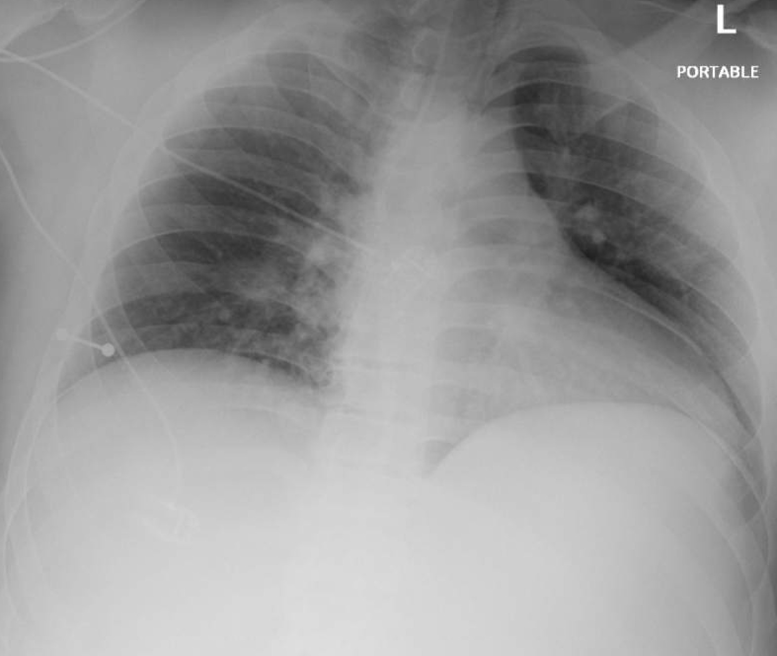
- Chest x-ray was obtained to evaluate for trauma which showed acute displaced left posterior 4th-7th rib fractures. No pleural effusion or pneumothorax was seen.
- After administration of ibuprofen, acetaminophen, and oxycodone, the patient’s pain was still reported as 8-9/10. The patient was offered and consented to an erector spinae plane block for multimodal pain control.
- The block was performed at bedside using ultrasound guidance (Figures 1-2). Half an hour after the block was performed, the patient’s pain had reduced to 4/10. The patient reported increased range of motion and subsequently was able to walk himself to the bathroom.
- The patient was subsequently cleared by trauma surgery for discharge home with multimodal pain control and follow up with PCP.


Discussion
Effective pain control in patients with rib fractures is critical to prevent complications such as atelectasis, pneumonia, and respiratory failure.1,2 Traditional management often relies on systemic opioids, which carry known risks including respiratory depression, cough suppression, and delirium.3 The erector spinae plane block (ESPB) is a regional anesthesia technique that provides effective analgesia for thoracic wall pain while reducing opioid requirements.4,5,6 In this case, ESPB resulted in a clinically meaningful reduction in pain and improved range of motion after failure of multimodal oral analgesia and facilitated safe discharge from the emergency department. The ESPB is well suited for the emergency room setting as it can easily be done at bedside using ultrasound guidance and is performed away from the pleura and other critical structures.5,6 Additionally, this technique is more technically straightforward in comparison to paravertebral or epidural blocks, which require greater technical expertise and carry higher risk.7,8 By targeting the fascial plane at the level of the transverse process, there is consistent blockade of the dorsal rami, with variable anterior spread to the ventral rami and intercostal nerves, allowing the ESPB to effectively provide broad unilateral analgesia across multiple rib levels4,5,9,10 (Figures 3-5).


References:
- Hamilton DL, Manickam B. Erector spinae plane block for pain relief in rib fractures. Br J Anaesth. 2017;118(3):474-475. doi:10.1093/bja/aex013
- Luftig J, Mantuani D, Herring AA, Dixon B, Clattenburg E, Nagdev A. Successful emergency pain control for posterior rib fractures with ultrasound-guided erector spinae plane block. Am J Emerg Med. 2018;36(8):1391-1396. doi:10.1016/j.ajem.2017.12.060
- Peek J, Smeeing DPJ, Hietbrink F, Houwert RM, Marsman M, de Jong MB. Comparison of analgesic interventions for traumatic rib fractures: a systematic review and meta-analysis. Eur J Trauma Emerg Surg. 2019;45(4):597-622. doi:10.1007/s00068-019-01116-w
- Forero M, Adhikary SD, Lopez H, Tsui C, Chin KJ. The erector spinae plane block: a novel analgesic technique in thoracic neuropathic pain. Reg Anesth Pain Med. 2016;41(5):621-627. doi:10.1097/AAP.0000000000000451
- Kumar G, Kumar Bhoi S, Sinha TP, Paul S. Erector spinae plane block for multiple rib fracture done by an emergency physician: a case series. Australas J Ultrasound Med. 2021;24(3):167-172. doi:10.1002/ajum.12261
- Jiang M, Peri V, Ou Yang B, Chang J, Hacking D. Erector spinae plane block as an analgesic intervention in acute rib fractures: a scoping review. Local Reg Anesth. 2023;16:81-90. doi:10.2147/LRA.S414056
- Palachick BJ, Carver RA, Byars DV, Martyak MT, Collins JN. Erector spinae plane blocks for traumatic rib fractures performed by nonspecialized emergency physicians: a prospective, interventional study. Am Surg. 2022;88(9):2124-2126. doi:10.1177/00031348221078428
- Elawamy A, Morsy MR, Ahmed MAY. Comparison of thoracic erector spinae plane block with thoracic paravertebral block for pain management in patients with unilateral multiple fractured ribs. Pain Physician. 2022;25(6):483-490.
- Ivanusic J, Konishi Y, Barrington MJ. A cadaveric study investigating the mechanism of action of erector spinae blockade. Reg Anesth Pain Med. 2018;43(6):567-571. doi:10.1097/AAP.0000000000000789
- Chin KJ, El-Boghdadly K. Mechanisms of action of the erector spinae plane (ESP) block: a narrative review. Can J Anaesth. 2021;68(3):387-408. doi:10.1007/s12630-020-01875-2


")


