Brandon Woo, Elaine Yu
A 28 year old male with unknown past medical history presented to the emergency department with a single gunshot wound sustained to the anterior chest just prior to arrival. He was brought in by family members who reported a witnessed traumatic gunshot wound to the chest by an unreported individual. The patient complained of trouble breathing and was severely agitated in the setting of pain. He required 75 mg IV ketamine for moderate sedation while obtaining vital signs, ultrasound, and intravenous access. The team prepared for urgent intubation in the setting of possible respiratory decompensation and activated massive transfusion protocol.
Vitals: BP 151/81 | Pulse 104 | Temp 97.8º F (36.6ºC) | Resp 27 | Wt 122.2 kg (269 lb 6.4 oz) | SpO2 93%
On physical exam, the patient was in acute distress and diaphoretic, reporting trouble breathing but speaking in full sentences. He was alert and oriented to person, place, and event. Primary trauma assessment: patent airway, rapid shallow breathing, and capillary refill less than 2 seconds. Secondary survey revealed an approximately 1 cm open wound with active bleeding to the subxiphoid region. There were no step-offs or deformities to the spine and no other obvious signs of trauma externally. There were equal breath sounds, and 2+ pulses were present in the radial and posterior tibialis arteries. The remainder of the exam was unremarkable.
A FAST exam was performed to evaluate for free fluid, hemothorax, or pericardial effusion.

Figure 1: Subxiphoid view

Figure 2: RUQ view

Figure 3: LUQ view

Figure 4: Pelvic view (transverse).

Figure 5: Pelvic view (longitudinal).
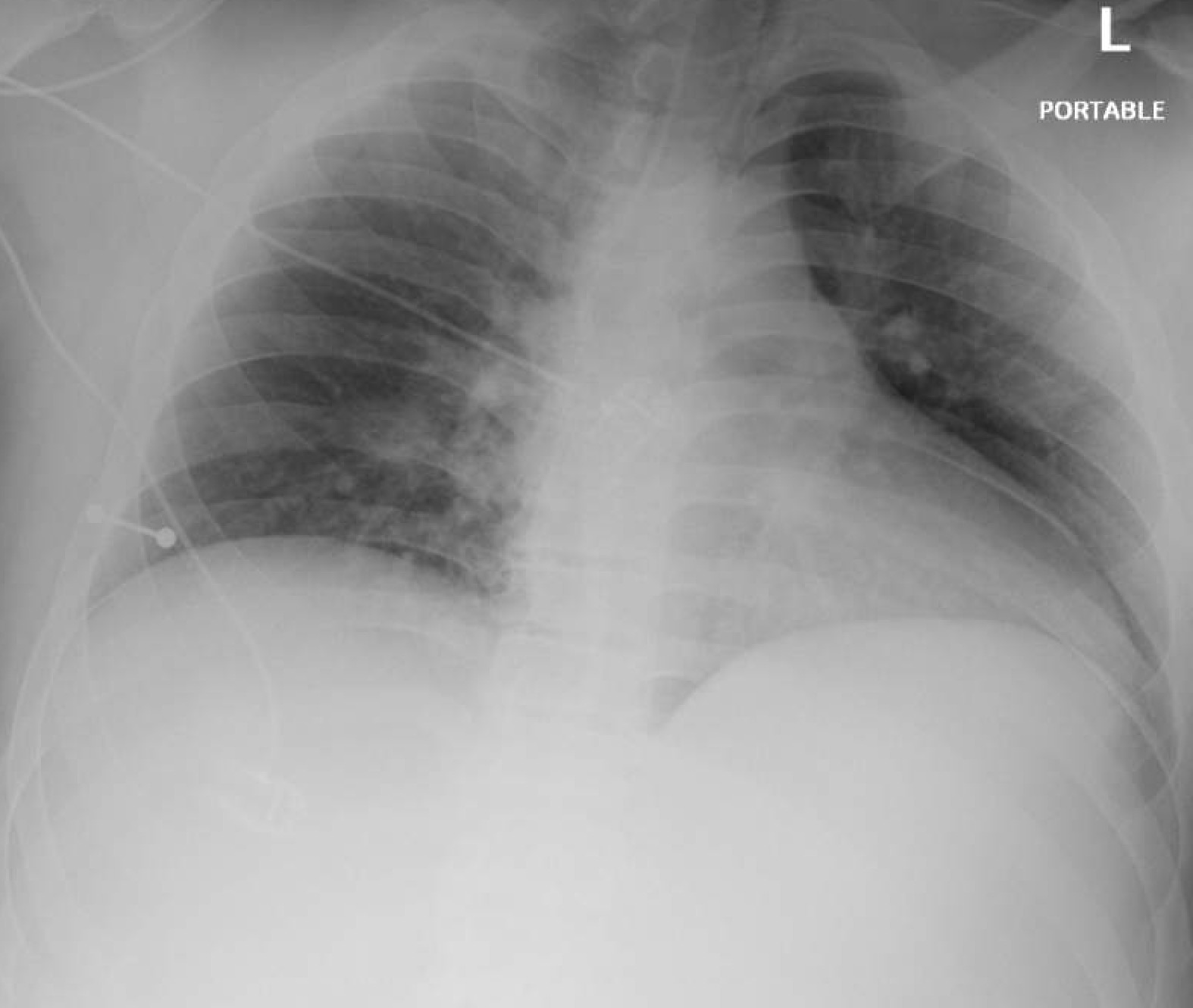
Figure 6: Chest X-Ray
Discussion:
The extended Focused Assessment with Sonography for Trauma (eFAST) is a validated rapid, bedside trauma tool, classically validated in hemodynamically unstable patients with blunt trauma where delays in imaging may be fatal. However its use in penetrating trauma is situational. The sensitivity of ultrasound is significantly lower for bowel injury, retroperitoneal bleeding, and intra-abdominal bleeding [1]. This patient was hemodynamically stable, which typically decreases the urgency for bedside ultrasound. However the patient’s agitation, tachypnea, and uncertain injury trajectory of a subxiphoid entry wound made thoracic and abdominal evaluation critical.
The presence of pelvic free fluid (Figures 4,5) and tachycardia with hyperdynamic ventricles (Figure 1) raised concern for intra-abdominal injury and potential hemorrhage. The location of free fluid in a supine trauma patient is influenced by gravity, with the rectovesical or rectouterine pouch being common sites of accumulation [1]. This patient had been upright prior to arrival and was agitated and repositioned multiple times, raising the possibility that the visualized pelvic fluid was gravitational rather than an isolated pelvic organ injury.
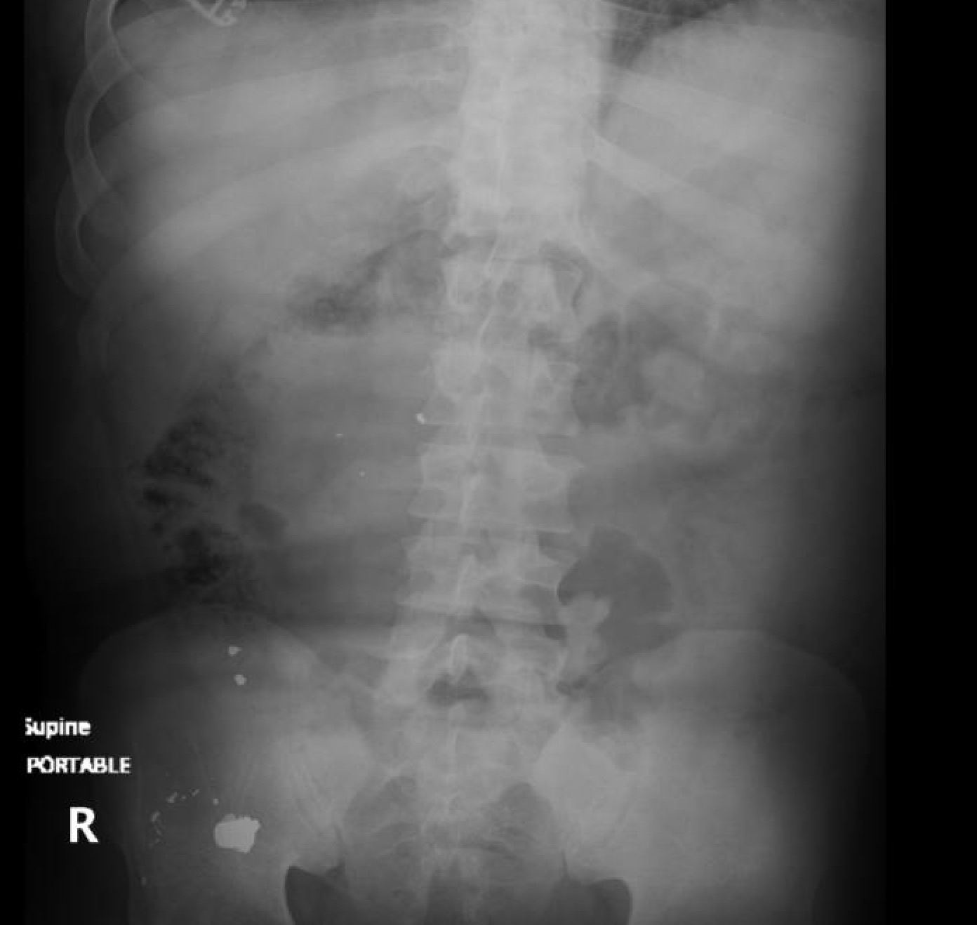
Figure 7: Abdominal X-ray.
Free fluid was visualized only in the suprapubic view, which may be due to localized pelvic injury, supported by the bullet location on KUB (Figure 7), or redistributed by gravity from another source, given that the patient had been upright and repositioned prior to evaluation. The absence of fluid in the right or left upper quadrants does not exclude intra-abdominal hemorrhage, particularly in the setting of penetrating trauma, where eFAST sensitivity varies significantly by region and injury type [1,2]. Retroperitoneal bleeding or bowel injury are often missed with eFAST alone, both possibilities in this case.
A meta analysis of over 24,000 trauma patients demonstrated eFAST sensitivity and specificity, respectively, for intra-abdominal free fluid (74%, 98%), pericardial effusion (91%, 94%), pneumothorax (69%, 99%) [2]. In the suprapubic view, sensitivity ranges from 80 - 90%, with specificity ~ 99%, and detection thresholds as low as 100 - 150 mL [2]. The RUQ view, Morison’s pouch, has a sensitivity 85 - 96% and specificity 98%, but typically requires at least 200 mL of fluid for reliable detection [2]. The LUQ view, splenodiaphragmatic space, demonstrates sensitivity 70 - 85% with specificity 98 - 99% [2]. These ranges reflect variations in body habitus, fluid pooling, and operator dependency. These views are widely taught per guidance of the ACEP Sonoguide [5].
A randomized controlled trial found that while FAST did not reduce mortality, it significantly improved workflow and led to a faster time to diagnosis, fewer CT scans, shorter hospital stays, and lower costs [3]. Additional studies have shown that even when eFAST yields false negative results, outcomes are favorable as long as patient status is closely monitored and reassessed [4]. These statistics demonstrate eFAST’s strength in ruling in injuries when positive, but its limitations in ruling out injuries when negative, especially in penetrating trauma, where sensitivity can range from 28-100% depending on trajectory, organ involved, and injury severity [5].
In this case, eFAST was used as an adjunct to clinical decision making, not a replacement for formal imaging or serial exam. Its findings enabled structured handoff, avoiding unnecessary invasive interventions, and highlighted how point-of-care ultrasound can be safely integrated into trauma workflows even when patients fall outside classical indications. Additional data are needed to clarify how patient position prior to arrival, upright versus supine, affects the sensitivity and specificity of eFAST in trauma assessment, particularly in the evaluation of intra-abdominal free fluid.
References:
- Tewari V, et al. The efficiency of focused assessment with sonography for trauma (FAST) vs. CT in penetrating torso trauma. J Emerg Trauma Shock. 2024;17(1):3–9.
- Netherton S, Milne WK, Proctor C, et al. Diagnostic accuracy of eFAST in trauma: a systematic review and meta-analysis. Can J Emerg Med. 2019;21(6):727–738.
- Stengel D, Bauwens K, Sehouli J, et al. Point-of-care ultrasonography for diagnosing thoracoabdominal injuries: a randomized controlled trial. JAMA. 2017;317(11):1110–1119.
- Sierzenski PR, et al. Clinical outcomes after false-negative FAST in hypotensive trauma patients. BMC Emerg Med. 2023;23:45.
- American College of Emergency Physicians (ACEP). Trauma Ultrasound (eFAST). Sonoguide. Available at: https://www.acep.org/sonoguide/basic/fast
- https://www.bcpocus.ca/organscans/efast/