The Predictive Value of Bedside Ultrasound to Restore Spontaneous Circulation in Patients with PEA: A Systematic Review and Meta-Analysis

Background

Cardiac arrest remains one of the leading causes of death in the United States and is frequently encountered in the emergency department (ED). It is defined as cessation of cardiac function and lack of circulation. Cardiopulmonary resuscitation (CPR) improves outcomes especially if it is performed within minutes of cardiac arrest. According to recent American Heart Association (AHA) statistics, approximately,  10.6% of patients who experience cardiac arrest survive to hospital discharge [1]. On the other hand, pulseless electrical activity (PEA) is a form of cardiac arrest in which patients continue to have organized cardiac electrical activity without a palpable pulse. This patient population's overall survival is much lower with 2.4% of patients surviving to hospital discharge [2]. Until recently, there has been an incomplete understanding of the the term PEA and what this means physiologically. With the advent of ultrasound (US), there has now been elucidation of two forms of PEA. True-PEA (tPEA) which lacks cardiac activity on US, has poor survival rates, while pseudo-PEA (pPEA) which demonstrates some cardiac activity on US,  shows improved survival,  potentially due to altering standard ACLS protocol driven management. The following study specifically looks at the data evaluating the predictive value of US in patients presenting in cardiac arrest with PEA.

The predictive value of bedside ultrasound to restore spontaneous circulation in patients with pulseless electrical activity: A systematic review and meta-analysis.

Clinical Question

Does bedside US predict the restoration of spontaneous circulation in patients with pulseless electrical activity?

Methods & Study Design

  • Design
    • Systematic review and meta-analysis
    • Data from MEDLINE, EMBASE, Cochrane library databases (inception to June 2017)
    • Statistical analysis
      • Review Manager 5.4 and Stata 12
      • I2 statistics to assess heterogeneity
      • Random effects model for all polled outcome measures
      • Begg’s test for publication bias
  • Study Eligibility Criteria
    • Adults with PEA
    • Cardiac US was used to detect cardiac activity
    • ROSC defined as primary outcome
    • Prospective/ observational studies
    • Written in English
    • 2x2 contingency table can be formed from data

Results

Included Studies

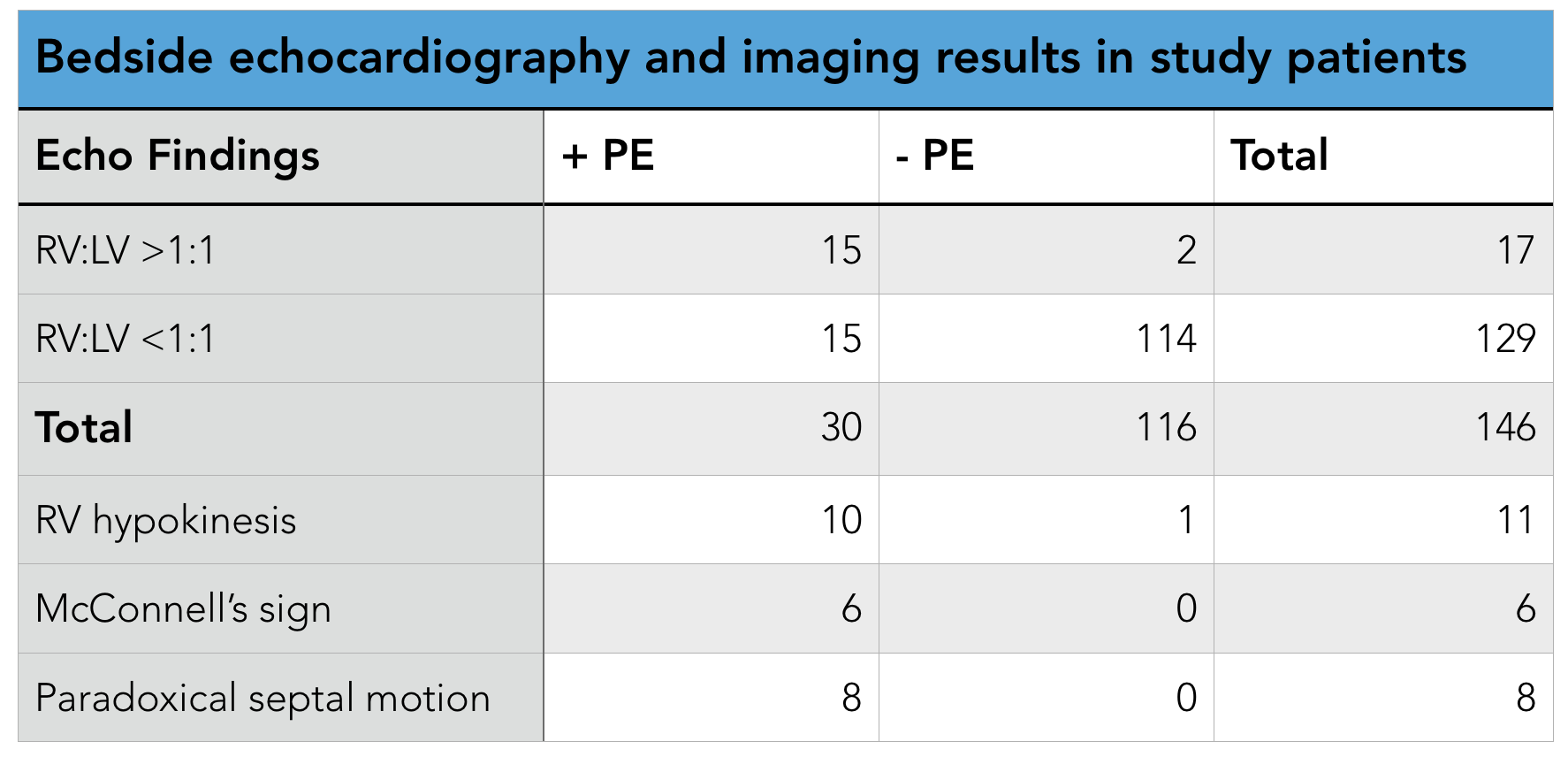
    • 11 studies with 777 patients with PEA included
    • 230 patients had ROSC
    • 42/343 "true-PEA" patients had ROSC
    • 188/434 "pseudo-PEA" patients had ROSC
    • Patients with pPEA were 4.35x more likely to experience ROSC than those tPEA (Risk ratio 4.35, confidence interval 2.20-8.63, p<0.00001, significant statistical heterogeneity I2= 60%)

Limitations

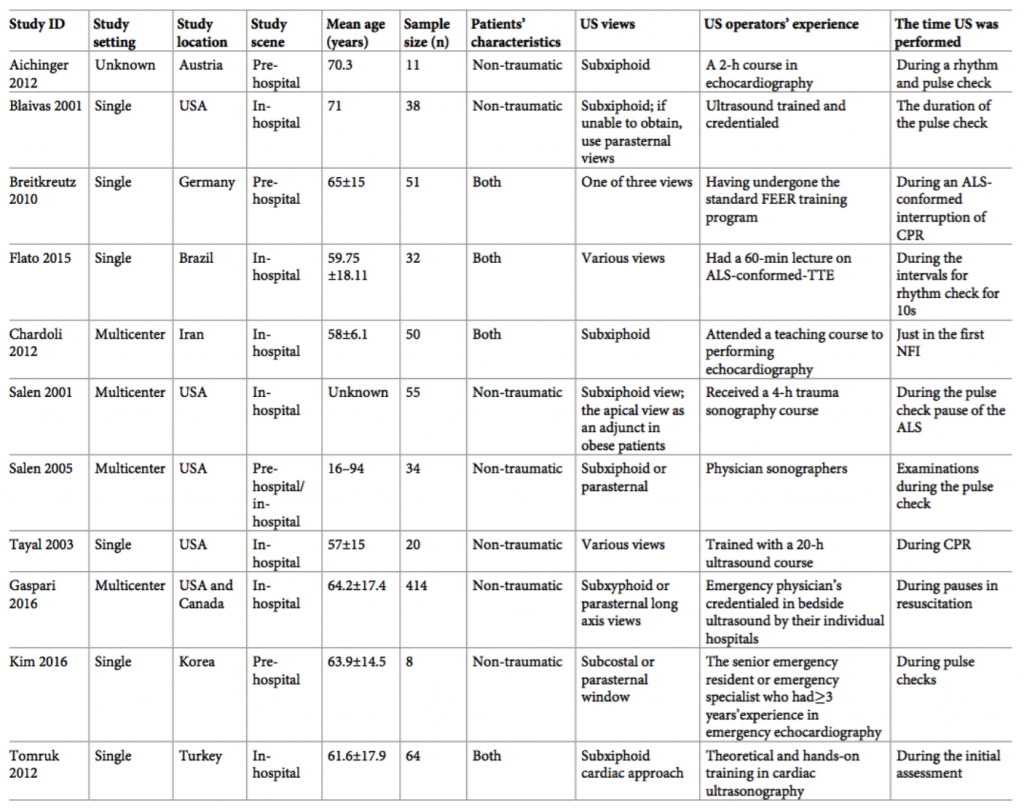
    • Significant heterogeneity amongst the 11 studies
      • 4 studies enrolled both trauma and non-trauma patients
      • In 3 studies, US evaluation occurred in the pre-hospital setting
    • Large confidence interval
    • Small pooled sample size
    • Varying protocols and US views used in different studies to determine cardiac activity
    • Varying definition of ROSC between studies

Authors Conclusion


"In cardiac arrest patients who present with PEA, bedside US has an important value in predicting ROSC. The presence of cardiac activity in PEA patients may encourage more aggressive resuscitation. Alternatively, the absence of cardiac activity under US could be promoted as a way of confirming a poor prognosis and used to support the decision to terminate resuscitative efforts."

Our Conclusion

This study found that patients in cardiac arrest with pPEA (i.e. cardiac motion on ultrasound) have higher ROSC than those with tPEA (i.e. no cardiac motion on ultrasound). The exact risk ratio for ROSC quoted in their results should be interpreted with caution since this meta-analysis included studies with vastly different characteristics. The 11 studies included took place in 9 different countries over the span of 15 years, included different US views (subxiphoid, parasternal), varied settings (pre-hospital and in-hospital US studies), varied patient populations (some studies included traumatic cardiac arrest) and had varying US operator experience. Additionally, other factors such as time to initiation of CPR, length of CPR, and the previous health of the patient were not accounted for. These limitations can affect the accuracy of the risk ratio presented in this study.  That being said, even with significant heterogeneity in this study, resulting in a very wide confidence interval, the lower limit of the risk ratio (2.20) still finds statistical significance for higher rate of ROSC in patients with pPEA compared to patients with tPEA.

This study essentially confirms what is already known from previous data (specifically the Gaspari study which represents the majority of patients in this meta-analysis) but fails to address the big question of "Does US guided resuscitation provide a mortality benefit in the management of cardiac arrest?" This is a complex question that takes into account multiple other questions including the debate over US increasing interruptions in chest compressions, the use of US to identify immediately reversible causes of cardiac arrest (i.e. tamponade, massive PE) , the true definition of cardiac standstill (which calls the results of all cardiac arrest studies thus far into questions), and ultimately, can US be used to determine if further resuscitation is futile? As with all advances in technology, finding the right niche to benefit the patient is of upmost importance and at this point in time, the utility of US in cardiac arrest remains to be determined.

The Bottom Line

Bedside ultrasound can be used to determine pPEA from tPEA in patients with cardiac arrest. This may help guide resuscitation efforts as patients with pPEA have increased rates of ROSC.

Authors

This post was written by Tina Vajdi, MS4 at UCSD. Review and further commentary was provided by Michael Macias, MD, Ultrasound Fellow at UCSD.

References

    1. Mozaffarian D, Benjamin EJ, Go AS, Arnett DK, Blaha MJ, Cushman M et al. Heart Disease and Stroke Statistics-2016 Update: A Report From the American Heart Association. Circulation 2016; 133(4):e38– e360. https://doi.org/10.1161/CIR.0000000000000350 PMID: 26673558
    2. Engdahl J, Bang A, Lindqvist J, Herlitz J. Factors affecting short- and long-term prognosis among 1069 patients with out-of-hospital cardiac arrest and pulseless electrical activity. Resuscitation 2001; 51 (1):17–25. PMID: 11719169
    3. Gaspari R, e. (2018). Emergency department point-of-care ultrasound in out-of-hospital and in-ED cardiac arrest. - PubMed - NCBI . Ncbi.nlm.nih.gov. Retrieved 20 April 2018, from https://www.ncbi.nlm.nih.gov/pubmed/27693280

Focused Transesophageal Echocardiography by Emergency Physicians is Feasible and Clinically Influential

Background

Cardiac ultrasound is frequently used in the emergency department (ED) to effectively identify  pericardial effusion, differentiate causes of shock, assess left ventricular function, and guide cardiopulmonary resuscitation (CPR). However, cardiac ultrasound employed in the ED is usually transthoracic echocardiography (TTE) as opposed to transesophageal echocardiography (TEE). TTE can often be limited, especially in critically ill patients and patients with high BMI. TEE offers the ability to reliably obtain continuous high-quality images that can be performed without interrupting CPR. Despite this, TEE is not often employed in the ED due to a variety of factors, including transducer cost, invasiveness, physician training, and hospital culture.

Focused Transesophageal Echocardiography by Emergency Physicians is Feasible and Clinically Influential: Observational Results from a Novel Ultrasound Program


Clinical Question

Is TEE performed by emergency medicine trained  physicians, in the emergency department setting, feasible and does it provide clinical utility?

Methods & Study Design

  • Design
    • Retrospective Review
  • Population
    • Study performed during a TEE in the ED pilot program by an academic emergency medicine program comprising 2 separate EDs, one of which is a regional trauma center.
    • All patients who underwent TEE in the ED during the 2-year program period were included.
  • Exclusion criteria
    • None
  • Intervention
    • TEE was performed on critically ill, intubated patients. Most commonly, the mid-esophageal 4-chamber view, followed by the transgastric short axis, mid-esophageal long axis, and bicaval views  were obtained.
  • OutcomesThe clinical impact of TEE, divided into two categories:
      • Diagnostic influence on clinical decision making
      • Therapeutic influence on procedures, medications, fluids, and CPR

Results

    • 54 TEE exams performed with 100% probe insertion success rate
      • 83% on first attempt
      • 11% required multiple attempts
      • 6% required use of a laryngoscope
      • 98% of exams produced images that were interpretable by the operator
    • TEE was diagnostically influential in 78% of cases
      • Excluded cardiac cause of arrest (56%)
      • Identified depressed left ventricular function (15%)
      • Identified hypovolemia (13%)
      • Identified regional wall motion abnormalities (6%)
      • Identified aortic dissection (4%)
    • TEE was therapeutically influential in 67% of cases
      • Influenced changes to CPR (43%)
      • Directed cessation of resuscitation (30%)
      • Guided hemodynamic support (26%)
    • No major adverse effects from probe placement identified

Strengths & Limitations

  • Strengths
    • TEE exams were performed successfully by 14 different emergency physicians at 2 separate sites after only 4 hours of training, which demonstrated well the feasibility of TEE use in the ED on a more widespread basis.
    • Well-described outcomes
  • Limitations
    • Retrospective
    • Relatively small sample size
    • No comparison with TTE

Author's Conclusions

“ED- based TEE showed a high degree of feasibility (98% determinate rate) and clinical utility, with a diagnostic and therapeutic influence seen in the majority of cases. Focused TEE demonstrates the most promise in patients who are intubated and have either undifferentiated shock or cardiac arrest.”

Our Conclusions

This study demonstrates that performing TEE in the ED is both feasible and safe, and can be implemented with limited training of the physician staff. It also shows that TEE does have some clinical utility in the ED, specifically the detection of aortic dissection. However, the most common therapeutic effect noted in the study was the assessment of CPR quality, which can typically be assessed with less invasive means such as femoral pulse palpation and waveform capnography. The other common findings noted in the study (i.e. depressed ejection fraction, hypovolemia, guidance of hemodynamic support) can typically be assessed with more traditional and less invasive TTE. In order to truly evaluate the utility of TEE in the ED, a prospective study showing a comparison of TEE with TTE, and other less invasive diagnostic modalities, would need to be performed. That being said, having the ability to diagnose aortic dissection at the bedside and to guide resuscitation via direct cardiac visualization during ongoing CPR are important considerations. This is a promising pilot study that opens up the door for further research evaluating the utility of TEE in the ED, however at this point, it is not clear whether it will perform better than traditional TTE and other clinical adjuncts in both diagnostic and therapeutic abilities.

The Bottom Line

Performing TEE in the ED is both feasible and safe, and does provide useful clinical information. However more studies are required in order to assess the true clinical utility of this modality.

Authors

This post was written by Toby Matt, MS4 at UCSD. It was reviewed by Michael Macias, MD, Ultrasound Fellow at UCSD.

References

    1. Arntfield R, e. (2017). Focused Transesophageal Echocardiography by Emergency Physicians is Feasible and Clinically Influential: Observational Results from a Novel Ultraso... - PubMed - NCBI . Ncbi.nlm.nih.gov. Retrieved 30 October 2017, from https://www.ncbi.nlm.nih.gov/pubmed/26508495

Variability in Interpretation of Cardiac Standstill Among Physician Sonographers

Background

The use of point-of-care echocardiography to inform termination or continuation of cardiopulmonary resuscitative efforts remains controversial [1,2]. Current understanding of its utility in prognostication is limited by varying definitions of cardiac activity. Definitions of cardiac standstill range from absence of “organized contractile activity (nonfibrillating) with a decrease in chamber size” to absence of “any visible movement of the myocardium, excluding movement of blood within the cardiac chambers or isolated valve movement” to absence of “any detected atrial, valvular, or ventricular motion within the heart” [3-5]. Without a consistent definition of cardiac standstill, it is difficult to interpret studies reporting conflicting resuscitation outcomes in cardiac arrest.

Variability in Interpretation of Cardiac Standstill Among Physician Sonographers

Clinical Question

What is the interrater reliability among providers in classification of cardiac standstill in point-of-care echocardiography?

Methods & Study Design

  • Design
    • Cross-sectional convenience sample survey
  • Population
    • Eligible: Residents, fellows, and faculty practicing in emergency medicine, critical care, or cardiology in attendance at one of six weekly emergency medicine (EM) conferences held at the following locations:
      • Icahn School of Medicine at Mount Sinai
      • Beth Israel Medical Center
      • St. Luke’s-Roosevelt Hospital
  • Exclusion criteria
    • Providers who had previously participated at a prior conference
  • Intervention
    • Participants were presented with the following clinical scenario: “55-year-old man in cardiac arrest who remains pulseless after 20 minutes of CPR”
    • Participants were shown 15 clips (6 seconds each, looped for 20 seconds total) presenting a variety of sonographic features
    • Asked to identify presence or absence of cardiac activity
    • Responses transmitted via remote polling devices
    • No definition of cardiac activity was provided
  • Outcomes
    • Primary: interrater reliability in interpreting cardiac standstill (Krippendorff’s alpha coefficient)
    • Secondary: subgroup analyses by specialty, training level, and self-described point of care (POC) ultrasound experience

Results

    • 127 participants (majority EM residents with basic ultrasound skills)
    • Overall moderate agreement with respect to identifying cardiac standstill (alpha 0.47)
    • Clips with stronger agreement:
      • No myocardial contraction
      • Myocardial contraction
      • Strong myocardial contraction
    • Clips with poorer agreement:
      • Valve flutter
      • Mechanical ventilation
      • Weak myocardial contraction
    • Moderate agreement across all training levels and self-reported ultrasonographic skill levels

Strengths & Limitations

  • Strengths
    • All participants saw the same clips
    • Response time limited (similar to clinical practice)
  • Limitations
    • Bias: recruitment from academic conferences
    • Majority with no or basic self-reported ultrasonographic skill level
    • Reported discussion among participants throughout survey

Author's Conclusions

“Our results support the possibility that previous studies have been subject to variability in the interpretation of cardiac standstill.”

Our Conclusions

We agree with the authors’ conclusions that there appears to be substantial variability in the interpretation of cardiac standstill. This study highlights a weakness in the current literature examining the utility of POC echocardiography used during resuscitation futility assessment. While this study does not provide data on clinical outcomes of standstill misclassification, it identifies a potential weakness in the available research. It is difficult to interpret studies reporting outcomes after standstill (such as meaningful survival) when the predictor is not consistently identified.

As a follow-up study, it would perhaps be interesting to see how the interrater reliability changes when participants are provided with a clear definition of cardiac standstill. Does the variability persist even with a uniform definition? If this improves interrater reliability it would provide additional support for the need for a consensus definition across future studies.

The Bottom Line

There is significant variability in classification of cardiac standstill among providers. A uniform definition of standstill may reduce this variability and aid in the interpretation of studies reporting conflicting outcomes after cardiac arrest.

Authors

This post was written by Carly Dougher, MS4 at UCSD. It was reviewed by Michael Macias, MD, Ultrasound Fellow at UCSD.

References

    1. Blyth, L., Atkinson, P., Gadd, K. & Lang, E. Bedside Focused Echocardiography as Predictor of Survival in Cardiac Arrest Patients: A Systematic Review: Echocardiography in Cardiac Arrest. Acad. Emerg. Med. 19, 1119–1126 (2012).
    2. Cohn, B. Does the Absence of Cardiac Activity on Ultrasonography Predict Failed Resuscitation in Cardiac Arrest? Ann. Emerg. Med. 62, 180–181 (2013).
    3. Schuster, K. M. et al. Pulseless Electrical Activity, Focused Abdominal Sonography for Trauma, and Cardiac Contractile Activity as Predictors of Survival After Trauma: J. Trauma Inj. Infect. Crit. Care 67, 1154–1157 (2009).
    4. Gaspari, R. et al. Emergency department point-of-care ultrasound in out-of-hospital and in-ED cardiac arrest. Resuscitation 109, 33–39 (2016).
    5. Kim, H. B., Suh, J. Y., Choi, J. H. & Cho, Y. S. Can serial focussed echocardiographic evaluation in life support (FEEL) predict resuscitation outcome or termination of resuscitation (TOR)? A pilot study. Resuscitation 101, 21–26 (2016).

Right ventricular dilatation on bedside echocardiography performed by emergency physicians aids in the diagnosis of pulmonary embolism

Background

Pulmonary embolism (PE) is a disease entity with a high mortality rate, ranging from 2.5-33%. Frequently, its diagnosis is delayed or frankly missed and often it is only discovered during autopsy. Around 66% of deaths occur during the first hour of presentation and 75% of deaths during the initial hospitalization. The mechanism of morbidity/mortality for PE is thought to be secondary to right ventricle (RV) outflow obstruction, leading to circulatory collapse. Delays in diagnosis have been linked to issues with imaging (wait times, schedules), contrast in the setting of renal impairment, and poor IV access.

In the emergency department, it is not only critical to identify patients with PE, but also to identify those who are at risk for decompensation and poor outcomes. This can be accomplished by evaluating for signs of RV dysfunction which has been associated with RV failure, hemodynamic collapse, and death. Previous studies have shown that right ventricular dysfunction has been found in 27-40% of normotensive patients with PE. Well studied markers of RV dysfunction include elevated biomarkers [1], specific ECG findings (RBBB, tachycardia, S1Q3T3, anterior TWI, ST elevation aVR, atrial fibrillation) [2], and RV dysfunction on echocardiography [3]. While biomarkers and ECG are readily available to emergency providers (EP), these are less specific for the diagnosis of PE and bedside echocardiography may prove to be more useful for evaluation of PE and RV dysfunction.

Right Ventricular Dilatation on Bedside Echocardiography Performed by Emergency Physicians Aids in the Diagnosis of Pulmonary Embolism

Clinical Question

Does evaluation for right ventricular dilation by emergency physicians using bedside echocardiography add diagnostic value in the evaluation for suspected pulmonary embolism? 

Methods & Study Design

  • Design
    • Prospective observational study
  • Population
    • Using a “convenience sample” population of patients who presented to the ED at Boston Medical Center from June 2009 – August 2011, with a moderate to high suspicion (pretest probability) of having a PE.  Wells score 2, those receiving PE imaging (CT, angio, V/Q scan), or those who came in with diagnosis of PE.
  • Exclusion criteria 
    • Non-english speakers
    • Prisoners
  • Intervention
    • Transthoracic echocardiography (blinded of confirmatory results) was performed by 4 ED docs, 1 with advanced training in cardiac sonography.  The other 3 had standard 1-month residency rotation in ultrasound and a minimum of 25 cardiac ultrasounds; plus, 10 hours hands-on and 10 hours image review with principal investigator.
    • Data collection
      • 3 views recorded: parasternal short & long axis, and apical 4-chamber, with primary measurement being qualitative assessment of RV size vs. LV size. Normal ratio (0.6:1)
        • Dilation defined as >1:1 RV:LV ratio
        • RV length and diameter or qualitative distension of RV apex adjacent to LV apex also assessed
      • They also recorded: RV function (nl vs. hypokinetic), paradoxical septal motion, and presence of McConnell’s sign.
      • All image reads were reviewed by the PI.
      • ED RAs then used chart review to compare findings to confirmatory imaging
        • PE was categorized as proximal vs distal
        • Disposition of patient was also documented 
  • Outcomes
    • Diagnostic characteristics
      • Sensitivity, specificity, PPV, NPV, positive and negative likelihood ratios
    • Presence of advanced signs of RV dysfunction
      • Right ventricular hypokinesis [qualitatively assessed as normal or hypokinetic], paradoxical septal motion, and McConnell’s sign

Results

    • Final analysis
      • 146 patients included in study
      • 126 with moderate pretest probability
      • 20 with high pretest probability
      • 126 with normal RV:LV ratio, 17 with increased RV:LV ratio
      • 30 had PE, of these 15 also had increased RV:LV ratio
    • Presence of RV dilation test characteristics
      • Sensitivity 50% (95% CI 32% to 68%)
      • Specificity 98% (95% CI 95% to 100%), a positive predictive value of 88%
      • Positive Predictive Value 88%  negative predictive value of 88%
      • Negative Predictive Value 88% (95% CI 83% to 94%).
      • Positive Likelihood Ratio 29 (95% CI 6.1% to 64%)
      • Negative Likelihood Ratio 0.51 (95% CI 0.4% to 0.7%)
      • Good observer agreement 96%, independent 100%

Strengths & Limitations

  • Strengths
    • Good concordance of sens/spec with prior study observations, although higher sensitivity
    • Good intra-observer agreement/reliability
  • Limitations
    • Single location, young population (less chronic diseases leading to RV changes)
    • Operator skill may not generalize to other physicians, other EDs.  PI was very experienced sonographer.
    • Convenience sample leading to possible selection bias.
    • Secondary outcomes under-powered.

Author's Conclusions

The authors conclude that right ventricular dilatation on bedside echocardiography may help emergency physicians rule in pulmonary embolism more rapidly by raising a provider’s index of suspicion before definitive testing. They also note that this evidence supports the concept that patients with a moderate to high pretest probability for pulmonary embolism and a bedside echocardiography result showing right ventricular dilatation should be considered for anticoagulation before definitive testing.

Lastly, they also comment on severity of PE, noting that patients with signs of advanced right ventricular dysfunction on bedside echocardiography (right ventricular dilatation with right ventricular hypokinesis, McConnell’ s sign, or paradoxical septal motion), tends to occur in patients with a larger clot burden who are more likely to be admitted to an ICU setting or have in hospital mortality (though this study was not powered appropriately for this analysis).

Our Conclusions

 We agree with the author's conclusions of this study that EP performed bedside echocardiography is a useful adjunct in the evaluation of suspected PE, both in identification of PE as well as risk stratification. We know that delays in diagnosis/treatment can lead to worse outcomes, however with the ability of EPs to perform bedside echocardiography and identify right ventricular dilation, this may reduce the time to both of these endpoints. It also seems reasonable that in patients who are moderate to high risk for PE, whom have evidence of right ventricular dilation on bedside echocardiography,  be empirically treated with anticoagulation prior to definitive imaging, with the caveat that they have no high bleed risk.

The Bottom Line

Emergency physician performed bedside echocardiography can be used reliably to increase provider's index of suspicion for PE in patients demonstrating RV dilation; however, given its poor sensitivity, it should not be used as a screening tool for PE.

Authors

This post was written by Hector Guerrero, MS4 at UCSD. It was reviewed by Michael Macias, MD, Ultrasound Fellow at UCSD.

References

    1. Weekes AJ, e. (2017). Diagnostic Accuracy of Right Ventricular Dysfunction Markers in Normotensive Emergency Department Patients With Acute Pulmonary Embolism. - PubMed - NCBI. Ncbi.nlm.nih.gov. Retrieved 25 September 2017, from https://www.ncbi.nlm.nih.gov/pubmed/26973178
    2. Shopp, J., Stewart, L., Emmett, T., & Kline, J. (2015). Findings From 12-lead Electrocardiography That Predict Circulatory Shock From Pulmonary Embolism: Systematic Review and Meta-analysis. Academic Emergency Medicine, 22(10), 1127-1137. doi:10.1111/acem.12769
    3. Dudzinski DM, e. (2017). Assessment of Right Ventricular Strain by Computed Tomography Versus Echocardiography in Acute Pulmonary Embolism. - PubMed - NCBI . Ncbi.nlm.nih.gov. Retrieved 25 September 2017, from https://www.ncbi.nlm.nih.gov/pubmed/27664798
    4. Dresden S, e. (2017). Right ventricular dilatation on bedside echocardiography performed by emergency physicians aids in the diagnosis of pulmonary embolism. - PubMed - NCBI . Ncbi.nlm.nih.gov. Retrieved 25 September 2017, from https://www.ncbi.nlm.nih.gov/pubmed/24075286

Ultrasound use during cardiopulmonary resuscitation is associated with delays in chest compressions

Background

Point-of-care ultrasound (POCUS) has quickly become a core adjunct used in the emergency department (ED) during cardiopulmonary resuscitation (CPR). Specifically, it is now frequently used to evaluate for reversible causes of cardiac arrest (i.e. cardiac tamponade, pulmonary embolism), guide resuscitation, and prognosticate on patient outcomes based on presenting cardiac activity [1]. However at this time, the benefits of the use of POCUS during CPR are not yet clear in terms of patient centered outcomes. What is known to have an effect on patient outcomes is providing high quality CPR, with minimal interruptions, and early defibrillation [2]. POCUS during CPR is often performed during pulse checks and it can take ample time to obtain sufficient views for proper interpretation. One concern is that this can result in unnecessary delays in resuming chest compressions during CPR, leading to harmful effects on patient outcomes.

Ultrasound use during cardiopulmonary resuscitation is associated with delays in chest compressions


Clinical Question

Does use of point-of-care ultrasound (POCUS) in cardiopulmonary resuscitation lengthen the duration of pulse checks beyond the guideline recommendation of 10 seconds?

Methods & Study Design

  • Design
    • Prospective cohort study
  • Population
    • This was a single center study performed at an adult, urban, tertiary care, academic medical center
    • All patients, 18 years of age or older, who presented to the emergency department (ED) in cardiac arrest or in whom cardiac arrest occurred while in the ED were eligible for enrollment
  • Exclusion criteria 
    • No documentation of a pulse check
    • Not placed in one of three designated resuscitation rooms with continuous video monitoring capability
    • Video monitoring obtained was not available or image quality was too poor for extraction of data
  • Intervention
    • All cardiac arrest resuscitations were recorded by video camera
      • Researchers recorded the duration of pulse checks (in milliseconds) and whether POCUS was used
  • Outcomes
    • Duration of pulse checks with the use of POCUS

Results

    • 23 patients enrolled
    • 123 individual rhythm checks
    • The use of POCUS significantly increased the pulse-check duration by 8.4 seconds (95% CI, 6.7-10.0 [p<0.0001])
    • Age and BMI did not affect the duration of pulse checks or CPR interruptions
    • No findings on any of the POCUS images prompted a procedure
    • Survival to ED discharge/hospital admission: 35% (8/23)
    • Survival to hospital discharge: 4% (1/23)
    • Survival at 30 days: 4% (1/23)

Strengths & Limitations

  • Strengths
    • Data collected directly from video which removes any bias on recall of events that occurred during cardiopulmonary resuscitation
  • Limitations
    • Small number of patients included in the study
    • Single center study reduces its external validity
    • No information of level of training of US operators
    • No commentary on the impact of POCUS on mortality or neurologic outcomes

Author's Conclusions

"The use of POCUS during cardiac arrest resuscitation was associated with an increase in the duration of pulse checks by 8.4 seconds, causing interruptions in high-quality chest compressions nearly double the 10-second duration recommended by current international cardiopulmonary resuscitation guidelines. It is critical for acute care providers to pay close attention to the duration of CPR interruptions when using POCUS during cardiac arrest resuscitation."

Our Conclusions

With the introduction of novel indications for POCUS, we need to be aware of both the benefits and harms. This is an important study that identifies a potential harm of POCUS during CPR. There is no doubt from this data that POCUS did result in prolonged pulse checks above the guideline recommended 10 seconds. While this study does not directly tell us that POCUS increases mortality in cardiac arrest (as it is much too small), there is clear evidence that increased interruptions in chest compressions during CPR leads to worse outcomes.

The utility of this study is that it identifies a problem with POCUS in which there are clear solutions. With this new evidence, we must take a thoughtful approach to use of POCUS during CPR. Some proposed solutions to address this problem include:

    • Identifying personnel during CPR who can verbalize the time spent during pulse check to make providers acutely aware of when CPR should be resumed ("10, 9, 8, 7...")
    • Recording a POCUS clip for 5 seconds, then reviewing the stored images while CPR is resumed
    • Having a care provider ready to perform POCUS exam with probe on chest just before chest compressions are withheld
    • Use of transesophageal (TEE) POCUS which allows for image acquisition during ongoing CPR

The findings of this paper are definitely not the end of POCUS in CPR, as I find that there are numerous reasons that it is useful (i.e. monitoring of proper chest compression location, identification of reversible etiology of cardiac arrest, prognostication, closure for health care providers during end of resuscitations...), however I would like to see larger studies on this topic as well as quality improvement and awareness of this potential harm of prolonged pulse checks secondary to POCUS utilization.

The Bottom Line

The use of POCUS during CPR may increase the duration of pulse checks beyond the recommended 10 seconds. Care providers should be aware of this potential harm and measures should be taken to prevent unnecessary delays in chest compressions.

Authors

This post was written by Michael Macias, MD, Ultrasound Fellow at UCSD.

References

    1. Gaspari R, e. (2017). Emergency department point-of-care ultrasound in out-of-hospital and in-ED cardiac arrest. - PubMed - NCBI . Ncbi.nlm.nih.gov. Retrieved 8 September 2017, from https://www.ncbi.nlm.nih.gov/pubmed/27693280
    2. Link MS et al. Part 7: Adult Advanced Cardiovascular Life Support: 2015 American Heart Association Guidelines Update for Cardiopulmonary Resuscitation and Emergency Cardiovascular Care. Circulation. 2015 Nov 3;132(18 Suppl 2):S444-64.

    3. Huis In 't Veld MA, e. (2017). Ultrasound use during cardiopulmonary resuscitation is associated with delays in chest compressions. - PubMed - NCBI . Ncbi.nlm.nih.gov. Retrieved 8 September 2017, from https://www.ncbi.nlm.nih.gov/pubmed/28754527

Feasibility and accuracy of bedside transthoracic echocardiography in diagnosis of acute proximal aortic dissection

Background

Acute aortic dissection is a life threatening condition that requires prompt diagnosis and definitive management; dissection involving the ascending aorta is undoubtably an indication for emergent surgical intervention. Previous data suggests that the mortality of type A dissection increases by 1-2% for every hour that passes which further highlights the importance of rapid diagnosis [1-2]. Currently, CT is considered the gold standard that enables the visualization of the entire aorta and can distinguish among the different types of acute aortic syndromes; however this is not always available, requires transferring patients to the CT scanner, and can ultimately generate a significant delay in treatment. Ultrasound is an easily available alternative imaging adjunct that may prove useful in rapid diagnosis of acute aortic dissection, specifically, type A dissection that require emergency surgical intervention.

Feasibility and Accuracy of Bedside Transthoracic Echocardiography in Diagnosis of Acute Proximal Aortic Dissection


Clinical Question

What is the accuracy of transthoracic echocardiography (TTE) in the diagnosis of acute type A aortic dissection in comparison to CT (with reference to the intra-operative diagnosis)?

Methods & Study Design

  • Design
    • Retrospective chart review
  • Population
    • This was a single center study involving patients  transferred due to suspected acute type A aortic dissection
    • Cardiac surgery for type A dissection was conducted in 172/178 patients (1 patient refused the operation and died, 5 patients underwent cardiac arrest and died prior to transfer to the operating room)
      • Because intra-operative findings were considered the gold standard reference for the presence of aortic dissection, the 6 patients who died without cardiac surgery were excluded from the final analysis
    • Inclusion criteria:
      • Referral for an urgent surgery due to proximal aortic dissection (Stanford classification Type A)
      • Available results of both CT and bedside TTE
    • Excluded
      • Patients who died prior to cardiac surgery
      • 1 patient who refused surgery
      • Patients who underwent surgical repair of acute type A aortic dissection based on TTE without confirmatory CT
  • Intervention
    • TTE was performed in the emergency department by an “experienced echocardiographer" to evaluate for: maximum ascending aorta diameter, presence of a dissection flap in the ascending aorta, left ventricular ejection fraction, pericardial effusion (and cardiac tamponade), aortic valve morphology and severity of aortic regurgitation
      • Echocardiographic findings were compared to CT findings and intra-operative findings were used as a gold standard
  • Outcomes
    • Identification of type A aortic dissection by TTE
    • Correlation of TTE measurements of maximum ascending aortic diameter with CT and intra-operative findings

Results

    • Statistical analysis with chi square test did not show any statistically significant differences between CT and TTE in the detection of proximal aortic dissection.
    • Additionally, echo revealed concomitant abnormalities (i.e. bicuspid aortic valve, AV calcifications, moderate/severe aortic incompetence, cardiac tamponade), which were all confirmed intra-operatively and influenced the treatment strategy (graft vs. valve-sparing surgery).
    • In patients with any aortic valve abnormalities (bicuspid aortic valve, AV calcifications, significant aortic regurgitation) procedure of choice was replacement by a composite graft (77.59% vs. 49.12%), whereas patients with normal aortic valves were significantly more likely to have the valve sparing surgery (50.88% vs. 22.41%)
    • There was a strong positive correlation between maximum diameter of the ascending aorta measured by TTE and CT (correlation coefficient 0.869)

Strengths & Limitations

  • Strengths
    • This was a feasibility study, and they used a population with known acute type A aortic dissection to determine if TTE could be used to provide both a rapid and reliable diagnosis in proximal aortic dissection
    • Gold standard was intra-operative findings
  • Limitations
    • Retrospective analysis, meaning that the diagnosis of aortic dissection has either already been made or was strongly suspected prior to initiating scanning; some may argue this may falsely increase the noted sensitivity/specificity of TTE
    • All patients who underwent cardiac surgery for acute proximal dissection based on TTE without CT verification (~30% patients at their institution) were excluded from the analysis
    • The TTE was performed by personnel trained in advanced echocardiography which may lower the sensitivity/specificity of these findings in the hands of less experienced operators

Author's Conclusions

"Our data confirm that TTE is a reliable method for diagnosis of proximal aortic dissection. TTE provides a reliable value of maximum diameter of the ascending aorta in comparison to both CT and direct intra-operative measurement. Moreover, TTE gives the additional information that influences the operative technique of choice and identifies the high-risk patients (cardiac tamponade, severe aortic dilatation, severe aortic regurgitation). Our retrospective analysis confirms the pivotal role of TTE in the evaluation of the patients with suspected proximal aortic dissection in emergency room setting."

Our Conclusions

Our conclusions are very similar to author findings on this paper. From the emergency department standpoint, we need the ability to distinguish sick patients from not sick patients and TTE in suspected acute aortic dissection does just that. Looking at this data, TTE measurements of maximum ascending aorta diameter correlate very well with intra-operative measurements. Furthermore, TTE is very accurate at identifying complications of type A aortic dissection such as decompensated heart failure (due to acute aortic regurgitation) and cardiac tamponade, both of which will alter surgical management.

What this means is that if you suspect aortic dissection, a bedside echo should be performed immediately looking for ascending aorta enlargement, dissection flap, and/or complications of dissection. If found, cardiac surgery can confidently be consulted and the patient can either be pushed to the operating room if unstable or pushed directly to the CT scanner by the emergency medicine provider. What this does not mean is that your work up stops here if no findings of dissection are found. If you are truly concerned about aortic dissection then the next step is to proceed with CT for definitive rule out. For more information on evaluation of acute aortic dissection, please read our recent case here.

The Bottom Line

The use of TTE in suspected proximal aortic dissection facilitates a rapid and reliable diagnosis, and shortens the delay to definitive treatment in a subset of high-risk patients.

Authors

This post was written by Ryan Shine, MS-4 at UCSD. It was edited by Michael Macias, MD.

References

    1. HIRST AE Jr, e. (2017). Dissecting aneurysm of the aorta: a review of 505 cases. - PubMed - NCBI Ncbi.nlm.nih.gov. Retrieved 26 August 2017, from https://www.ncbi.nlm.nih.gov/pubmed/13577293

    2. Hagan PG, e. (2017). The International Registry of Acute Aortic Dissection (IRAD): new insights into an old disease. - PubMed - NCBI Ncbi.nlm.nih.gov. Retrieved 26 August 2017, from https://www.ncbi.nlm.nih.gov/pubmed/10685714

    3. Sobczyk, D., & Nycz, K. (2015). Feasibility and accuracy of bedside transthoracic echocardiography in diagnosis of acute proximal aortic dissection. Cardiovascular Ultrasound, 13(1). doi:10.1186/s12947-015-0008-5

Case # 4: To Bolus or Not to Bolus?


A 67 year old male with a PMHx of DM presents with a chief complaint of cough and generalized weakness.

Vitals: T 102.4 HR 127 BP 77/58  RR 24 O2 88% on RA

You place the patient on O2 via nasal cannula and activate the sepsis protocol. He is empirically treated with broad spectrum antibiotics and IVFs are started. The chest x-ray shows multifocal pneumonia and you call hospital medicine to admit the patient.  “What’s the blood pressure now,” the hospitalist asks. You glance at the monitor and murmur back, “92/63, but he looks pretty good.” The hospitalist asks you to insert a central line, start vasopressors, and contact the ICU. Instead, you wheel the ultrasound machine into his room, and ultrasound his IVC. Does this patient require a central line and vasopressors?

Answer and Learning Point

Answer

No, the patient’s IVC is small and collapsing almost 75% with normal respiratory variation. This predicts a fluid-responsive state. The patient was given another liter of lactated ringers, his blood pressure improved to 108/69, and his lactate cleared. You start maintenance IV fluids, call the hospitalist back, and the patient is admitted upstairs and does well.

Learning Points

    • Fluid responsiveness is a controversial topic that continues to plague emergency medicine physicians and intensivists alike
    • In patients whom a fluid bolus is being considered, ultrasound can be a useful tool to assess for cardiac function, lung fluid status (interstitial edema) and whether a patient will improve their cardiac output in response to this fluid challenge
    • A recent study showed that the cIVC (inferior vena cava collapsibility) can be used as a predictor of who will be a fluid responder [1]
      • cIVC = (IVC expiratory diameter - IVC inspiratory diameter)/IVC expiratory diameter
      • Patients with a cIVC > 25% are likely to be fluid responders (LR + 4.56)
      • Patients with a cIVC < 25% are unlikely to be fluid responders (LR - 0.16)
    • The IVC should be examined in the subxiphoid region with the probe in a sagittal plane, and can be found by first identifying the right atrium and following this caudally
      • A back-up approach involves using the liver as an acoustic window , placing the probe in the mid axillary line in a coronal plane,  and fanning anteriorly and posteriorly until the IVC is visualized
      • The IVC should be measured 3 cm caudal to the junction of the right atrium and IVC [2]
    • M-mode can be used to evaluate the cIVC and has the advantage of measuring the exact same spot along the IVC over an extended period of time
    • As with all adjuncts to clinical decision making, fluid responsiveness should not be determined solely on a single ultrasound measurement such as cIVC but should be taken into context with the rest of the clinical picture

Author

This post was written by Amir Aminlari, MD, Ultrasound Fellowship Director at UCSD.

References

Corl KA, e. (2017). Inferior vena cava collapsibility detects fluid responsiveness among spontaneously breathing critically-ill patients. - PubMed - NCBI . Ncbi.nlm.nih.gov. Retrieved 19 August 2017, from https://www.ncbi.nlm.nih.gov/pubmed/28525778

Nagdev AD, e. (2017). Emergency department bedside ultrasonographic measurement of the caval index for noninvasive determination of low central venous pressure. - PubMed - NCBI . Ncbi.nlm.nih.gov. Retrieved 19 August 2017, from https://www.ncbi.nlm.nih.gov/pubmed/19556029

Diagnosing Acute Heart Failure in the Emergency Department: A Systematic Review and Meta-analysis

Background

The chief complaint of “dyspnea” represents a very large cohort of patient who present to the emergency department. While acute heart failure (AHF) is a very common diagnosis in the setting of dyspnea, the diagnosis remains challenging when the emergency physician is presented with the undifferentiated dyspneic patient. Interestingly, emergency physicians have varied approaches to the work up and diagnosis of these patients and it is not clear as to which diagnostic element is most crucial in confirming the diagnosis of AHF. Spoiler: Ultrasound proves to be quite useful.

Diagnosing Acute Heart Failure in the Emergency Department: A Systematic Review and Meta-analysis‌


Clinical Question

What are the operating characteristics of the diagnostic elements available to the emergency physician for diagnosing acute heart failure?

Methods & Study Design

  • Design
    • This is a systematic review evaluating index test operating characteristics in diagnosing AHF. A medical literature search was performed using PubMed and EMBASE, evaluating peer-reviewed published papers from 1965 through 2015
    • Individual systematic reviews for each index test were conducted by two separate physicians and thereafter reconciled to obtain a comprehensive set of studies on the topic. These were then screened against the inclusion/exclusion criteria for final inclusion into the meta-analysis
    • The reference standard used was a final diagnosis of AHF based on review of clinical data by independent reviewers who were blinded to the study’s primary index test
  • Population
    • All studies included involved patients presenting to the emergency department (ED) with the chief complaint of “dyspnea.”
  • Outcomes
    • Pooled sensitivities, specificities and likelihood ratios (LRs) of index tests for diagnosing acute heart failure in patients presenting to the ED with dyspnea
    • They specifically looked at the following index tests in evaluation of AHF: history and physical exam, ECG, chest x-ray, BNP and NT-ProBNP, lung ultrasound (US), and bedside echocardiography
  • Excluded
    • Patients presenting to urgent care with dyspnea
    • Patients with chronic, compensated heart failure
    • Studies focusing on prognosis or therapeutics and not the diagnosis of AHF
    • Studies with ultrasound images that were not obtained and interpreted by emergency physicians

Results

    • History and Physical: S3 most specific finding for AHF (+LR 4)
    • ECG: Found to be insensitive and unspecific for diagnosing or ruling out AHF
    • CXR: Pulmonary edema was the most specific finding (LR + 4.8). All other imaging findings were insensitive for ruling out heart failure
    • BNP and NT-Pro-BNP: Quite sensitive for ruling out AHF at a threshold of 100/300pg/dL
    • Lung Ultrasound:
      • Presence of >3 B-lines in >2 lung fields is very specific for the presence of AHF
      • Lack of this also sensitive for ruling out acute heart failure
      • High inter-rater reliability
    • Bedside Echo
      • ED provider evaluation of systolic function had high inter-rater reliability with the ultimate ejection fraction assessed by cardiologists on formal echo
      • Restricted Mitral Inflow very specific for ruling in diastolic AHF in patients with preserved systolic function

Strengths & Limitations

  • Strengths
    • Very thorough analysis of the operating characteristics of a plethora of diagnostic elements and sub-elements available to the emergency physician in diagnosing AHF
    • Authors didn’t exclude comorbidities, etiology of AHF or if there was an underlying arrhythmia which increased the generalizability of their results
    • All data was screened and evaluated by two separate physicians
  • Limitations
    • The reference standard was a final diagnosis of AHF made by physicians in retrospective fashion which weakens this as a “gold standard.”
    • The authors did not specifically evaluate or reconcile whether the heart failure was left or right sided
    • Each of the tests or test characteristics were assessed in isolation to determine the likelihood of heart failure. The likelihood of AHF when considering multiple index tests was not assessed
    • As in all large meta-analyses, some spectrum bias may exist as inclusion/exclusion criteria varied among included studies. However, ome of this heterogeneity is likely countered by the pooled analysis and is unlikely to drastically change the calculated LRs

Author's Conclusions

"Bedside lung US and echocardiography appear to the most useful tests for affirming the presence of AHF while natriuretic peptides are valuable in excluding the diagnosis."

Our Conclusions

This is one of the most thorough studies available to assess the likelihood that a patient presenting to the emergency department has acute heart failure based on an index test. There are many old standby diagnostic modalities available to emergency physicians including the history, physical exam, and chest x-ray. Unfortunately, these diagnostic elements are relatively non-specific in establishing that a patient’s acute symptoms are likely or unlikely due to heart failure. BNP and NT Pro BNP are quite useful in ruling out heart failure however these tests take time to result. Ultrasound is rapidly becoming a fundamental tool in every emergency physicians tool belt and should be utilized alongside the primary patient assessment in determining the likelihood that a patient has heart failure. The presence or absence of B lines (>3 in at least 2 fields) is quite specific for ruling in heart failure and the absence is nearly as sensitive as a normal BNP or NT Pro BNP, too. And in the event of systolic heart failure, echo is a great modality to rapidly assess a patient’s pump function; our interpretation is consistent with the formal result obtained by cardiologists [2].

Caution must nevertheless be maintained when evaluating these results. The LRs found in these studies were calculated independently of other findings and in reality, the emergency physician takes multiple factors from the history, physical exam, and other diagnostic modalities, to ultimately come to a definitive diagnosis. Essentially, the short answer is that no single test should be taken as definitive in diagnosis of AHF (or any diagnosis for that matter) and the emergency physician should follow a bayesian approach using pre- and post- test probabilities from their fund of knowledge to rule in and rule out cannot miss diagnoses. Lastly, with regards to this study, physicians should be wary about interpreting these results in the context of renal failure primarily but also superimposed pneumonia or underlying concern for pulmonary embolism as these patients were excluded in a number of papers included in this meta-analysis.

The Bottom Line

Bedside ultrasound to evaluate for the presence or absence of pulmonary edema should be an integral part of the emergency physicians approach to evaluating patients to the emergency department with undifferentiated dyspnea.

Authors

This post was written by Matt Correia, MD PGY-2 at UCSD. It was edited by Michael Macias, MD.

References

    1. Martindale JL, e. (2017). Diagnosing Acute Heart Failure in the Emergency Department: A Systematic Review and Meta-analysis. - PubMed - NCBI . Ncbi.nlm.nih.gov. Retrieved 14 August 2017, from https://www.ncbi.nlm.nih.gov/pubmed/26910112
    2. Moore CL, e. (2017). Determination of left ventricular function by emergency physician echocardiography of hypotensive patients. - PubMed - NCBI . Ncbi.nlm.nih.gov. Retrieved 14 August 2017, from https://www.ncbi.nlm.nih.gov/pubmed/11874773

Case # 2: A Needle In the Haystack

A 40 year old male presented with 3 days of progressive dyspnea on exertion. He notes he was in a normal state of health prior to this and played basketball daily without issue but now he can no longer walk across the room without becoming winded. He has no chest pain, a normal chest x-ray and an ECG demonstrating sinus tachycardia

Vitals: HR 109 BP 110/72 RR 22 O2 96

ezgif.com-optimize+(2)

Answer and Learning Point

Answer

There is evidence of severe aortic regurgitation and aortic root dilation (~6 cm) on this parasternal long axis view. In a patient without any previous cardiac history with new aortic regurgitation this is concerning for acute aortic dissection. Cardiac surgery was consulted immediately and the patient was taken straight to CT scan for confirmation of type A aortic dissection. The patient was in the OR within 1 hour and had an excellent outcome.

Learning Point

Aortic dissection is quite uncommon (~5-30 per 1 million people per year) and is often seen in patients with chronic uncontrolled hypertension or other diseases such as bicuspid aortic valve, Marfan Syndrome or Ehlers-Danlos Syndrome. Unfortunately all the "classic" indicators of dissection are actually not that common [1].  Traditionally we are taught that patients with acute aortic dissection will arrive hypertensive, while in actuality up to 1 in 4 patients with Stanford Type A dissection will have a presenting systolic blood pressure below 100 mmHg. Additionally, it is taught that a dissection presents as a “ripping or tearing” pain going to the back.  Looking at the data, while over 90% of patients felt that it was the worst pain they had ever experienced, only 50% of subjects described their pain as ripping or tearing (62% described pain as sharp), only 35% had any posterior chest pain, and only 85.4% of patients described the onset of their pain as ‘acute.’ [1]

The varied presentation of this disease makes aortic dissection difficult to diagnose, and the clinician should have a high index of suspicion for this life-threatening disease process.  This is where ultrasound comes in. Anyone who has a concerning chest pain story, pain above and below the diaphragm, chest pain + a neurological symptom, or signs and symptoms of acute heart failure without any previous cardiac history, should have a bedside ultrasound performed.  While ultrasound cannot rule out aortic dissection, it can rapidly identify complications of dissection and expedite care in these patients whom time is of the essence.

The Approach

Perform standard abdominal aorta ultrasound evaluating for aneurysm or intimal flap. Be sure to evaluate from proximal aorta, in the epigastric region, distally to the iliac vessels. A normal aorta caliber is < 3 cm.

Obtain a parasternal long axis view:

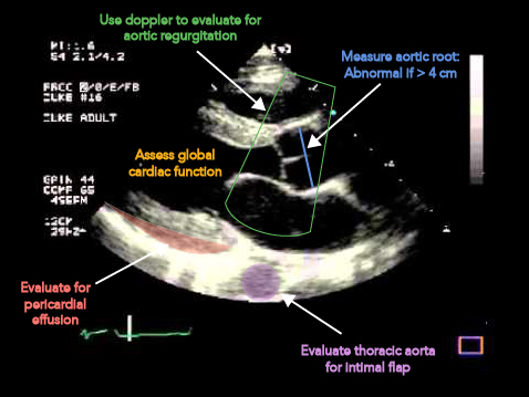
Measure aortic root, this should be less than 4 cm. There are varying opinions on where the best place to take this measurement is, I suggest measuring the largest area you see as it is better to be on the conservative side.
Apply color doppler to evaluate for aortic regurgitation.
Assess global cardiac function. This is useful to see if a patient is compensated or decompensated as well as assist with fluid/pressor management if needed.
Evaluate for pericardial effusion. If there is evidence of effusion and concern for Type A aortic dissection, this suggests that there is communication with pericardial sac.
Evaluate descending thoracic aorta for intimal flap

References

  1. Hagan PG, Nienaber CA, Isselbacher EM, et al. The International Registry of Acute Aortic Dissection (IRAD): New Insights Into an Old Disease. JAMA. 2000;283(7):897-903. doi:10.1001/jama.283.7.897.
  2. Taylor RA, e. (2017). Point-of-care focused cardiac ultrasound for the assessment of thoracic aortic dimensions, dilation, and aneurysmal disease. - PubMed - NCBI Ncbi.nlm.nih.gov. Retrieved 22 July 2017, from https://www.ncbi.nlm.nih.gov/pubmed/?term=22288871
  3. C, K. (2017). Emergency department diagnosis of aortic dissection by bedside transabdominal ultrasound. - PubMed - NCBI Ncbi.nlm.nih.gov. Retrieved 22 July 2017, from https://www.ncbi.nlm.nih.gov/pubmed/19549013
  4. Lang R, Bierig M, Devereux R, et al. Recommendations for Chamber Quantification: A Report from the American Society of Echocardiography’s Guidelines and Standards Committee and the Chamber Quantification Writing Group, Developed in Conjunction with the European Association of Echocardiography, a Branch of the European Society of Cardiology. J Am Soc Echocardiogr 2005; 18: 1440-63.
  5. Rubano E, e. (2017). Systematic review: emergency department bedside ultrasonography for diagnosing suspected abdominal aortic aneurysm. - PubMed - NCBI Ncbi.nlm.nih.gov. Retrieved 22 July 2017, from https://www.ncbi.nlm.nih.gov/pubmed/?term=23406071

Case # 1: The Acutely Winded Traveler

A 65 year old female presents with shortness of breath after a return flight from the Gold Coast of Australia to the United States.

Vitals: HR 107 BP 110/80 RR 22 O2 95

RV+Strain_Mead

Answer and Learning Point

Answer

There is right ventricular dysfunction demonstrated as septal bowing appreciated on this parasternal short axis view. This is concerning for a pulmonary embolism in the setting of the provided clinical context.

Learning Point

Echocardiography can be a useful adjunct to laboratory markers (i.e. BNP and troponin) and CTA for evaluation of right heart strain in normotensive patients presenting with concern for pulmonary embolism.  While there is building evidence that many patients presenting with pulmonary embolism are safe for discharge [1] , those patients that have evidence of right ventricular dysfunction are at higher risk for morbidity and mortality and may also be candidates for more advanced therapies, other than simple anticoagulation, such as catheter directed thrombolysis.  The most up-to-date evidence supports that emergency physicians can accurately perform echocardiography at the bedside to risk stratify patients presenting with concern for pulmonary embolism. In a recent study by Weekes et al, emergency physicians (EP) performed goal directed echocardiography to assess for right ventricular dysfunction. If any of the following criteria below were present, a patient was considered positive by goal directed echocardiography for right ventricular dysfunction:

This study found the EP goal-directed echocardiography sensitivity and specificity for right ventricular dysfunction to be 100% (CI 87% to 100%) and 99% (95% CI 94% to 100%), respectively [2]. Our patient ended up having a saddle embolus and underwent catheter directed thrombolysis and did well.

References

  1. Aujesky D, e. (2017). Outpatient versus inpatient treatment for patients with acute pulmonary embolism: an international, open-label, randomised, non-inferiority trial. - PubMed - NCBI . Ncbi.nlm.nih.gov. Retrieved 8 July 2017, from https://www.ncbi.nlm.nih.gov/pubmed/21703676

  2. Weekes AJ, e. (2017). Diagnostic Accuracy of Right Ventricular Dysfunction Markers in Normotensive Emergency Department Patients With Acute Pulmonary Embolism. - PubMed - NCBI . Ncbi.nlm.nih.gov. Retrieved 8 July 2017, from https://www.ncbi.nlm.nih.gov/pubmed/?term=26973178

Translate »