Caudal Edge of the Liver in the Right Upper Quadrant (RUQ) View Is the Most Sensitive Area for Free Fluid on the FAST Exam

ruq free fluid

Background

The FAST exam is a useful tool in screening for the presence of intraperitoneal free fluid in the setting of trauma. The utilization of ultrasound provides rapid imaging in the trauma bay that can help guide clinical decision making and the necessity for surgical intervention. The FAST exam is comprised of subxiphoid, right upper quadrant, left upper quadrant, and suprapubic views by ultrasound. Previous research has indicated that the RUQ, specifically the hepato-renal space (Morrison’s pouch), is the preferred area for the detection of free fluid.1,2 However, scarce research into the sub-divisions of each view has been performed.

Caudal Edge of the Liver in the Right Upper Quadrant (RUQ) View Is the Most Sensitive Area for Free Fluid on the FAST Exam

Clinical Question

The aim of this study was to determine what specific sub-divided areas of each FAST view were the most sensitive in the detection of intraperitoneal free fluid. 

Methods & Study Design

• Design 

This was a retrospective cohort analysis. 

• Population 

All patients who received a FAST exam at a single Level 1 trauma center over an 18-month period.

• Intervention 

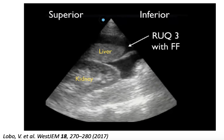
The RUQ, LUQ, and suprapubic views of the FAST were each subdivided into three additional sections for analysis. Specifically, the RUQ was divided into the hepato-diaphragmatic space (RUQ1), hepato-renal space (Morrison’s pouch) (RUQ2), and the caudal liver tip (RUQ3). The LUQ was divided into the spleno-diaphragmatic space (LUQ1), the spleno-renal space (LUQ2), and the inferior pole of the kidney (LUQ3). The suprapubic area was divided into the lateral sides of the bladder (SP1), posterior bladder and anterior pelvic organ space (SP2), and posterior uterus (in female subjects) (SP3). The subxiphoid view was excluded as the study was only interested in intraperitoneal free fluid.  

• Outcomes  

Each sub-quadrant of all positive FAST exams was analyzed for the presence of free fluid. 

Results

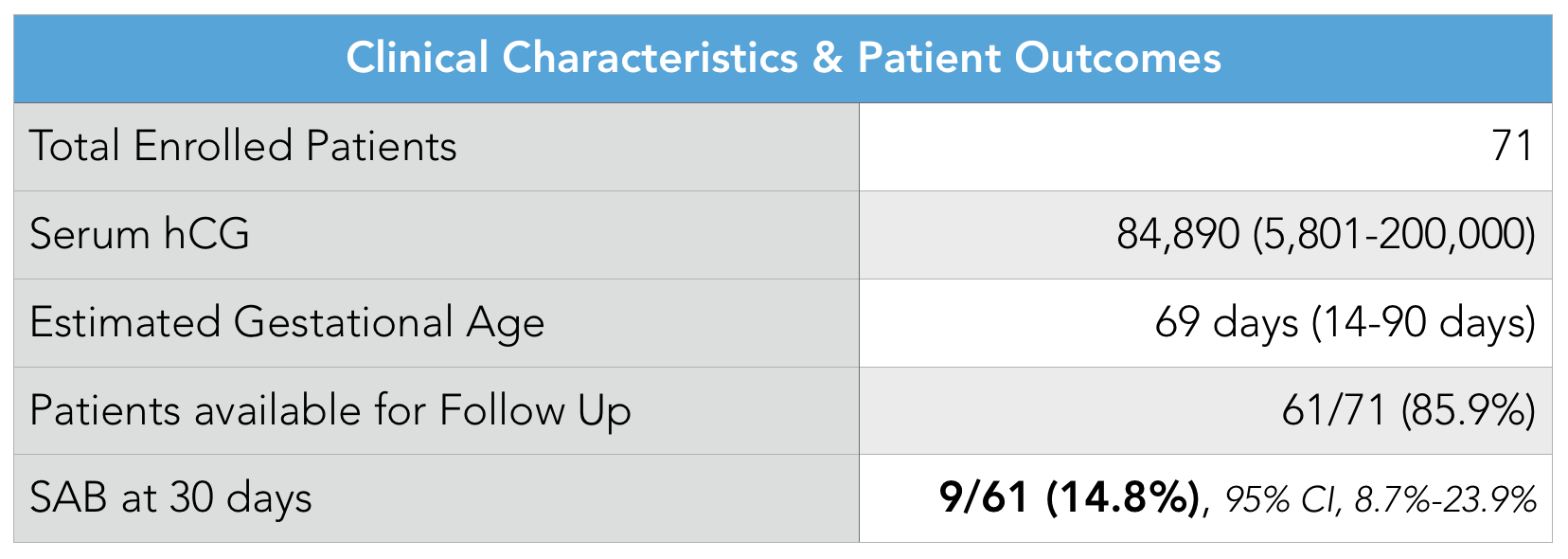
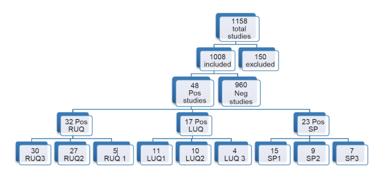
  • Of the 1,008 FAST scans included in the study, 48 (4.8%) were positive for free fluid. These findings were either confirmed by CT or intraoperatively. 
  • Of the positive FAST exams, 32 (66.7%) were positive in the RUQ, 17 (35.4%) were positive in the LUQ, and 23 (47.9%) were positive in the suprapubic region. 
  • Of the positive RUQ scans, 30 (93.8%) were positive in RUQ3, 27 (84.4%) in RUQ2, and 5 (15.6%) in RUQ1. 
  • Of LUQ scans, 11 (64.7%) were positive in LUQ1, 10 (58.8%) in LUQ2, and 4 (23.5%) in LUQ3. 
  • In the SP view, 15 (64.7%) were positive in SP1, 9 (58.8%) in SP2, and 7/9 (77.7%) in SP3.
  • No correlation was found between quadrants. FAST exam quadrants
  • ruq free fluid

Strength & Limitations

This was a simple, well done study that provides useful information on the FAST exam. The study featured a relatively small sample size of 48 positive FAST exams. There is a potential that in the time between a FAST exam and further intervention (CT or OR) further bleeding could have occurred, thus lowering the sensitivity of the FAST. The authors struggled to make conclusions regarding the suprapubic view between sexes due to a small sample size of positive SP views. 

Authors Conclusion

The caudal tip of the liver (RUQ3) is the most sensitive area for the detection of free fluid on FAST exam.


Our Conclusion

The FAST exam can be an extremely useful tool at the bedside or in the setting of trauma. All areas of the FAST should be properly viewed, with particular emphasis on the caudal tip of the liver. It is important to note that many FAST exams only showed free fluid in one area of one quadrant while showing no free fluid elsewhere, therefore it is important to assess all three areas in every view to increase the sensitivity of the FAST exam. 

The Bottom Line 

Despite previous emphasis on Morrison’s pouch, the caudal liver tip is a more sensitive indicator of intraperitoneal free fluid and should be properly visualized on every FAST exam.

Authors

This post was written by Oliver Marigold, MS4 at UCSD School of Medicine, Charles Murchison MD and Amir Aminlari MD.

References

  1. Von Kuenssberg Jehle, D., Stiller, G. & Wagner, D. Sensitivity in detecting free intraperitoneal fluid with the pelvic views of the FAST exam. The American Journal of Emergency Medicine 21, 476–478 (2003).
  2. AIUM Practice Guideline for the Performance of the Focused Assessment With Sonography for Trauma (FAST) Examination. Journal of Ultrasound in Medicine 33, 2047–2056 (2014).
  3. Lobo, V. et al. Caudal Edge of the Liver in the Right Upper Quadrant (RUQ) View Is the Most Sensitive Area for Free Fluid on the FAST Exam. WestJEM 18, 270–280 (2017).

Can Fluid Accumulation on Ultrasound Diagnose Necrotizing Fasciitis?

necrotizing fasciitis

Background

Necrotizing fasciitis (NF) is rapidly progressing, severe soft tissue infection with a mortality rate of 19.3% with treatment and significantly higher without treatment (1). Early diagnosis is essential to prompt surgical intervention and reduce morbidity and mortality. However, treatment can often be delayed because no laboratory or imaging test can definitively diagnose NF. Contrast-enhanced CT shows the best accuracy, but again is not perfect and can be difficult to obtain in unstable patients. MRI is similarly accurate, but even less feasible in the Emergency Department. Ultimately, it remains a surgical diagnosis.

Ultrasonography is a rapid, bedside, and non-invasive tool that has potential to accelerate assessment of patient with clinical suspicion for NF. There are ultrasonographic findings associated with NF diagnosis, including irregularity or thickening of deep fascia, subcutaneous emphysema, and fluid accumulation along the deep fascial plane (2-13). Considering this condition’s rapid progression, ultrasonography may enable physicians to quickly gauge disease severity and triage accordingly, prompting earlier surgery and bettering patient outcomes.

The Relationship Between Fluid Accumulation in Ultrasonography and Diagnosis and Prognosis of Patients with Necrotizing Fasciitis


Clinical Question

What is the relationship between ultrasonographic finding of fluid accumulation along the deep fascia and diagnosis and prognosis of necrotizing fasciitis?

What ultrasonographic findings are significantly different between NF patients and non-NF patients?

What is the ultrasonographic-detected depth of fluid accumulation along the deep fascia that offers the greatest accuracy to diagnosis of NF?

Is there a difference in the prognosis between NF patients with fluid accumulation compared to NF patients without fluid accumulation? 

Methods & Study Design

• Design 

Retrospective study with prospective enrollment

• Population 

This study was conducted at Chang Gung Memorial Hospital, a suburban academic tertiary care hospital.

Inclusion criteria: patients who visited the ED from February 2015 – November 2016 with clinical suspicion of NF of limbs based on symptoms and clinical signs (severe pain out of proportion, skin findings, rapid progression, crepitus, skin bullae, necrosis, or ecchymosis).

NF group: discharge diagnosis of NF, confirmed by pathology report showing necrosis after surgical intervention

Non-NF group: did not have surgical intervention or whose pathology report did not support NF diagnosis

Exclusion criteria: patients with ED visits between 24:00 – 7:00, non-lesion side also has fluid accumulation, age <18yo, prior antibiotics or debridement, lesions involving trunk area

• Intervention 

Ultrasonographic exam within 1 hour after ED arrival completed by one of three experienced emergency physicians who received an 8-hour basic and soft-tissue ultrasonographic training before the study

Orthopedic consult for surgical opinion

• Outcomes  

  • Diagnostic markers: irregularity or thickening of deep fascia, fluid accumulation, subcutaneous emphysema, subcutaneous cobblestone
  • Reasonable cutoff value of fluid accumulation along deep fascial plane for diagnosing NF according to receiving operating characteristic (ROC) curve
  • Prognostic markers: length of stay (LOS) in hospital, mortality, amputations, number of operations

Results

Ultrasound finding of fluid accumulation and irregular or thickened fascial layer were significantly different between NF and non-NF groups. All patients who had subcutaneous emphysema were in the NF group.

The best cutoff point of fluid accumulation to diagnose NF was 2mm, which had the best accuracy (72.7%), with sensitivity of 75%, a specificity of 70.2%, a positive predictive value of 71.7% and a negative predictive value of 72.7%.

NF patients with fluid accumulation had longer length of stay than NF patients without fluid accumulation (average: 39 days vs. 23 days). Number of operations were not significantly different between NF patients with and without fluid accumulation. All NF patients who had an amputation or died had fluid accumulation.


Overall mortality between NF and non-NF groups showed no significant difference.

Strength & Limitations

Strengths

  • Sample size was larger than other studies investigating ultrasonographic findings for NF diagnosis.
  • Study had a comparator groups with clear definitions (NF vs. non-NF).
  • Ultrasound training was standardized and assessed with inter-rater reliability between three emergency physicians as 100%.

Limitations

  • Small, imbalanced sample of NF patients for sensitivity and specificity analysis of fluid accumulation for amputation and mortality. 
  • Study excluded patients with truncal soft tissue infections.
  • Study excluded patients with prior antibiotics or debridement, which may have been NF patients with higher severity and worse prognosis.
  • Patient population were from south Taiwan exclusively.
  • NF patients had higher prevalence of specific co-morbidities (diabetes mellitus, liver cirrhosis, and alcohol use disorder), which could be confounding. 

Authors Conclusion

“The ultrasonographic finding of fluid accumulation along the deep fascia with a cutoff point of more than 2 mm of depth may aid in diagnosing NF. For the prognosis of NF, when fluid accumulation was present along deep fascia on ultrasound, patients with NF had longer lengths of hospital stays and were at risk of amputation or mortality. Ultrasonography is a point-of-care imaging tool that facilitates the diagnosis and prognosis of NF.” (14)

Our Conclusion

Consistent with prior studies and case reports (2-13), this study supports the role of ultrasound in the diagnosis of NF. Trained emergency physicians were able to successfully use ultrasound to detect significant imaging differences in NF patients, including fascial irregularity and deep fascial fluid accumulation. In comparison to Yen et al., this study suggests an even lower cutoff point of fluid accumulation along the deep fascia (2mm vs 4mm) for the highest diagnostic accuracy. We would caution that the finding of "fluid accumulation" was somewhat difficult to interpret in their study.

Further studies with larger sample sizes need to be completed. However, with the diagnostic and prognostic trends seen in this study, ultrasound should be considered as a timely, efficient imaging modality that can help identify patients with clinical suspicion of NF and accelerate OR intervention.

The Bottom Line 

Ultrasound is a viable imaging modality for patients with clinical suspicion of NF that could potentially expedite surgical intervention, though imaging findings may not be as easy to interpret as the authors lay out.

Authors

This post was written by Caresse Vuong, Charles Murchison MD and Amir Aminlari MD.

References

  1. Khamnuan P, Chongruksut W, Jearwattanakanok K, Patumanond J, Yodluangfun S, Tantraworasin A. Necrotizing fasciitis: Risk factors of mortality. Risk Manag Healthc Policy 2015;8:1–7.
  2. Castleberg E, Jenson N, Am Dinh V. Diagnosis of necrotizing fasciitis with bedside ultrasound: The STAFF exam. West J Emerg Med 2014;15:111–113.
  3. Tsai CC, Lai CS, Yu ML, Chou CK, Lin SD. Early diagnosis of necrotizing fasciitis by utilization of ultrasonography. Kaohsiung J Med Sci 1996;12:235–240.
  4. Wronski M, Slodkowski M, Cebulski W, Karkocha D, Krasnodebski IW. Necrotizing fasciitis: Early sonographic diagnosis. J Clin Ultrasound 2011;39:236–239.
  5. Yen ZS, Wang HP, Ma HM, Chen SC, Chen WJ. Ultrasonographic screening of clinically-suspected necrotizing fasciitis. Acad Emerg Med 2002;9:1448–1451.
  6. Bernardi, Emanuele, Antonello Iacobucci, Letizia Barutta, Elisa Pizzolato, Virna Olocco, and Bruno Tartaglino. “A-Lines in Necrotizing Fasciitis of the Lower Limb.” Journal of Ultrasound in Medicine 33, no. 11 (2014): 2044–46. 
  7. Chao, H. C., M. S. Kong, and T. Y. Lin. “Diagnosis of Necrotizing Fasciitis in Children.” Journal of Ultrasound in Medicine: Official Journal of the American Institute of Ultrasound in Medicine 18, no. 4 (April 1999): 277–81. 
  8. Hosek, William T., and Timothy C. Laeger. “Early Diagnosis of Necrotizing Fasciitis with Soft Tissue Ultrasound.” Academic Emergency Medicine 16, no. 10 (2009): 1033–1033. 
  9. Oelze, Lindsay, Stanley Wu, and Jennifer Carnell. “Emergency Ultrasonography for the Early Diagnosis of Necrotizing Fasciitis: A Case Series from the ED.” The American Journal of Emergency Medicine 31, no. 3 (March 1, 2013): 632.e5-632.e7. 
  10. Kehrl, Thompson. “Point-of-Care Ultrasound Diagnosis of Necrotizing Fasciitis Missed by Computed Tomography and Magnetic Resonance Imaging.” The Journal of Emergency Medicine 47, no. 2 (August 2014): 172–75. 
  11. Shyy, William, Roneesha S. Knight, Ruth Goldstein, Eric D. Isaacs, and Nathan A. Teismann. “Sonographic Findings in Necrotizing Fasciitis.” Journal of Ultrasound in Medicine 35, no. 10 (2016): 2273–77. 
  12. Hanif, Muhammad A., and Michael J. Bradley. “Sonographic Findings of Necrotizing Fasciitis in the Breast.” Journal of Clinical Ultrasound: JCU 36, no. 8 (October 2008): 517–19. 
  13. Valle Alonso, Joaquín, Ganapathiram Lakshmanan, and Yasser Saleem. “Use of POCUS Ultrasound in Sepsis, Bedside Diagnosis of Necrotizing Fasciitis.” QJM: An International Journal of Medicine 110, no. 10 (October 1, 2017): 687–88. 
  14. Lin, Chun-Nan, Cheng-Ting Hsiao, Chia-Peng Chang, Tsung-Yu Huang, Kuang-Yu Hsiao, Yi-Chuan Chen, and Wen-Chih Fann. “The Relationship Between Fluid Accumulation in Ultrasonography and the Diagnosis and Prognosis of Patients with Necrotizing Fasciitis.” Ultrasound in Medicine & Biology 45, no. 7 (2019): 1545–50. 


Bedside Ultrasound Identification of Infectious Flexor Tenosynovitis in the Emergency Department

flexor tenosynovitis

Background

Infectious flexor tenosynovitis (FTS) is a surgical emergency. If not treated promptly, infectious FTS carries significant morbidity including loss of function of fingers, necrosis of the tendon, and even digit amputation (1).

Infection can be caused in three ways: direct inoculation, contiguous spread, or hematogenous spread, as seen in cases of disseminated gonococcal infection. Tenosynovitis occurs when fluid collects between the visceral and parietal layer of the tendon, the most common location being in the hand and wrist.

Traditionally, diagnosis of infectious FTS is centered on the tetrad known as Kanavel’s signs (swelling of the finger, finger held in partially flexed position, pain on palpation of the flexor tendon, and pain on passive extension of the finger). While Kanavel’s signs are specific for infectious FTD, in a study of 41 participants with infectious FTS, only 54% of patients taken to the operating room (OR) had all of these signs (2). The gold standard of diagnosis remains surgical exploration and drainage. MRI can aid in the diagnosis of FTS, but this is rarely available in the ED. While radiographs may be obtained to look for trauma, osteomyelitis or a foreign body, they offer minimal to no additional benefit in diagnosing infectious FTS.   

This article presents a case of a 58-year-old man where point of care ultrasound (POCUS) identified tissue necrosis and fluid along the flexor tendon sheath of the hand, aiding in the rapid diagnosis of FTS, adding to the limited body of literature supporting use of POCUS for early diagnosis of infectious FTS.

Bedside Ultrasound Identification of Infectious Flexor Tenosynovitis in the Emergency Department

Clinical Question

Can point of care ultrasound be used in the emergency department to diagnose infectious FTS?

Methods & Study Design

• Design 

Case report.

• Population 

58 year old male with hypertension, diabetes and end stage renal disease.

• Intervention 

POCUS looking for fluid in flexor tendon sheath. Appropriate technique is shown in the image below, with a linear ultrasound probe placed on the palmar side of the wrist crease. Common findings of FTS are hypoechoic or anechoic fluid surrounding the flexor tendons.

ultrasound flexor tendons
Padrez et al. West J Emerg Med 2015, 16(2)

• Outcomes  

Accurate diagnosis of FTS


Results

The physicians found a moderate amount of fluid and echogenic material within the tendon sheath, as noted in the image below. Orthopedics was consulted and patient was started on broad spectrum antibiotics and taken to the operating room. They found extensive pus within the flexor tendon sheath and cultures grew Staph aureus


ultrasound flexor tenosynovitis
Padrez et al. West J Emerg Med 2015, 16(2)

Strength & Limitations

The POCUS exam the authors describe is practical and useful. This could feasibly be performed by clinicians with relatively little ultrasound training. As mentioned, FTS is a surgical emergency and remains largely a clinical diagnosis, so any modality that helps bring more certainty to the diagnosis, and lead to quicker definitive treatment, is welcome.

It is unclear what the level of ultrasound training was for the physicians who performed this exam. Another note, it may be difficult to distinguish rheumatologic from infectious causes of tenosynovitis using ultrasound, so clinical context is always important. They also mentioned this can only aid in increasing your suspicion for FTS, it cannot be used to rule it out. 

Authors Conclusion

POCUS may be an ideal adjunct for the ED physician in the evaluation of a patient with suspected infectious FTS 

Our Conclusion

We agree with the authors conclusions that POCUS can be a useful adjunct to clinical exam in diagnosing FTS, with the understanding that POCUS cannot rule out FTS or distinguish rheumatologic from infectious process.

The Bottom Line 

POCUS can be a useful adjunct to clinical exam in diagnosing FTS. Use the linear probe and place at the palmar side of the wrist crease, look for hypoechoic or anechoic material around the flexor tendons with possible thickening of the tendon itself. 

Authors

This post was written by Betial Asmerom and Amir Aminlari MD. Edited by Charles Murchison MD.

References

  1. Mamane, W. et al. Infectious flexor hand tenosynovitis: State of knowledge. A study of 120 cases. J. Orthop. 15, 701–706 (2018).
  2.  
  3. Hubbard, D., Joing, S. & Smith, S. W. Pyogenic Flexor Tenosynovitis by Point-of-care Ultrasound in the Emergency Department. Clin. Pract. Cases Emerg. Med. 2, 235–240 (2018).
  4.  
  5. Padrez et al. Bedsound Ultrasound Identification of Infectious Flexor Tenosynovitis in the Emergency Department. West J Emerg 2015. 16 (2). 


Ultrasound-guided supra-inguinal fascia iliaca block: a cadaveric evaluation of a novel approach

fascia iliaca block

Background

Regional anesthesia of the fascia iliaca is well-documented to be a successful way to control acute pain from hip fractures in the emergency department, particularly in those patients at high risk of complications from repeated doses of IV opioids. However, the majority of existing descriptions on technique for performing fascia iliaca blocks focus on approaching from inferior to the inguinal ligament, relying on supra-inguinal spread to block the lateral femoral cutaneous nerve in the iliac fossa. This study aims to investigate the utility in performing suprainguinal injection of anesthetic agents directly into the iliac fossa to provide regional anesthesia.

Ultrasound-guided supra-inguinal fascia iliaca block: a cadaveric evaluation of a novel approach

Clinical Question

Does injecting dye superior to the inguinal ligament provide significant uptake of dye in the femoral, LCFN, and ilioinguinal nerves in cadaveric models?

Methods & Study Design

• Design 

This is an informational study made to illustrate the utility of an alternative supra-inguinal approach for providing regional anesthesia in those patients presenting with acute hip or knee pain.

• Population 

Bilateral injections of 20 mL of 0.25% aniline blue dye were administered to six unembalmed cadavers, for a total of 12 injections available for analysis.

• Intervention 

Bilateral injections of 20 mL of 0.25% aniline blue dye were administered to six unembalmed cadavers via an ultrasound guided approach, and dye uptake was analyzed in anatomic nerve distributions.

Steps of superior approach to fascia iliaca block:

  • Place sterile probe over the inguinal ligament, close to the anterior superior iliac spine
  • Orient linear probe in parasagittal oblique orientation (superior aspect facing medially)
  • Rock the probe so beam faces laterally to enhance fascia iliaca
  • Move probe infero-medially along the line of inguinal ligament until femoral artery is imaged
  • Moving probe supero-laterally helps identify anterior inferior iliac spine (site of rectus femoris attachment)
  • As you move laterally, you see “rising” of ilium towards transducer
  • Identify deep circumflex artery 1-2cm superficial to fascia iliaca
  • Needle introduced 2-4cm inferior to inguinal ligament, and advanced through the fascia iliaca at the level of inguinal ligament.
  • “pop” as needle passes through fascial iliaca and into the iliacus muscle
  • Needle withdrawn to the fascia, and position confirmed by 1cc of local anesthetic.
  • Injection of fluid produces a“lens”that appears. The fluid should then hydro-dissect as it migrates into the iliac fossa
  • End point is when local anesthetic passes freely superiorly over the iliacus muscle and into the iliac fossa.

• Outcomes  

Dye uptake in the femoral, lateral femoral cutaneous nerve, and ilioninguinal nerves after supra-inguinal injection.

Results

    • There was extensive spread of dye identified in the iliac fossa. (50 to 144mm of spread)
    • 10 out of 12 injections also resulted in spread into the thigh along the femoral nerve
    • The femoral nerve was surrounded by dye in all cases
    • The LFCN was identified bilaterally in 5 cadavers, but one cadaver lacked both LFCN. LCFN was surrounded by dye in all cases in which it was present.
    • It is important to realize that the ilioinguinal nerve has significant anatomic variation. The ilioinguinal nerve passed over iliac crest onto iliacus muscle and re-emerged into the abdominal wall anteriorly in 8 cases. In the other four cases it remained in the abdominal wall. It was stained blue 7 out of 8 times in this procedure as it passed over iliac crest.

Strength & Limitations

    • There was no comparison group in this study (supra-inguinal approach was used in all cases).
    • There were significant anatomic variations between cadavers. One cadaver was found to have no lateral femoral cutaneous nerve. There were also significant anatomic variations in positioning of the ilioinguinal nerve.
    • Throughout the article, there were multiple references that the authors institution has performed over 150 supra-inguinal fascia iliata blocks without any short term complications, however no retrospective data was available in the article to confirm this.
    • Low sample population (6 cadavers, 12 nerve blocks)

Authors Conclusion

“Our cadaveric dye-injection study confirms that the ultrasound-guided supra-inguinal approach result in significant spread of injectate with simultaneous involvement of both the femoral nerve and LFCN, in the iliac fossa, in the all the cadavers in which we identified theses nerves by dissection.”

Our Conclusion

This article outlines an interesting alternative approach to providing regional anesthesia for acute pain control of the hip or knee. The majority of existing descriptions on technique for performing fascia iliaca blocks focus on approaching from inferior to the inguinal ligament, relying on supra-inguinal spread to block the lateral femoral cutaneous nerve in the iliac fossa. This study demonstrates consistent bathing of the LFCN and femoral nerve with dye in cadaveric subjects with a supra-inguinal approach. However, this study does not directly demonstrate superior efficacy of the suprainguinal approach when compared to infrainguinal fascia iliaca block. Further patient-oriented studies would be needed to make such a suggestion.

The Bottom Line 

Ultrasound guided suprainguinal fascia iliaca injection of dye in cadaveric subjects shows consistent and significant uptake of injectate of the femoral nerve and LFCN in the iliac fossa. Further studies are needed to show if this provides improved analgesia as compared to the conventional infrainguinal approach. 

Authors

This post was written by Casey Smith, MD. Review and further commentary was provided by Danika Brodak, MD, Emergency Ultrasound Fellow at UCSD and Amir Aminlari, MD, Ultrasound Faculty at UCSD.

References

  1. Hebbard P, Ivanusic J, Sha S. Ultrasound-guided supra-inguinal fascia iliaca block: a cadaveric evaluation of a novel approach. Anaesthesia. 2011;66(4):300‐305. doi:10.1111/j.1365-2044.2011.06628.x


Point-of-Care Ultrasonography for Evaluation of Acute Dyspnea in the ED

Background

Dyspnea is a common presenting symptom in the emergency department, and early diagnosis of underlying disease pathology is crucial in rapid intervention and treatment. Laboratory and radiological tests aid in the diagnosis, but often these results take time.1-3 Additionally, chest radiographs and chest CTs, the most common radiological tests in the evaluation of dyspnea, have several disadvantages including radiation risks and high costs. Unlike these modalities, point-of-care ultrasound (PoCUS) is cheap with no radiation risk, highly accurate, and has better sensitivity in detecting pneumothorax, pneumonia, and pleural effusions than CXR.4-7 In addition to being accurate and reliable, PoCUS can be performed rapidly to aid in early diagnosis and treatment of patients.

Point-of-Care Ultrasonography for Evaluation of Acute Dyspnea in the ED

Clinical Question

What is the feasibility and diagnostic accuracy of PoCUS for the management of acute dyspnea in the ED?

Methods & Study Design

  • Design:

Prospective, blinded, observational study

  • Population:

This study was conducted at Careggi University Hospital, a university-affiliated teaching hospital.

  • Inclusion Criteria:

Patients over the age of 18 with acute dyspnea of any degree. 

  • Exclusion Criteria:

Patients with dyspnea of traumatic origin, and those that were discharged from the emergency department after evaluation. 

  • Intervention:

All patients were primarily assessed by 2 separate emergency physicians with vital signs, history, physical exam, and EKG.

One physician performed a Lung, Cardiac, and IVC PoCUS.

One physician performed a standard workup using any combination of Chest X-Ray, Chest CT, Echocardiogram, labs, or Arterial Blood Gas.

Both physicians were asked to make up to 2 diagnoses based on their results.

Possible diagnoses: Heart Failure, Acute Coronary Syndrome, Pneumonia, Pleural Effusion, Pericardial Effusion, COPD/asthma, Pulmonary Embolism, Pneumothorax, ARDS/ALI, Other.

  • Outcomes

Primary: 

Accuracy of diagnosis:

Follow-up chart review determined the reference diagnosis. Results were compared to the diagnosis obtained from the ultrasound group and the standard workup group.

Secondary: 

Time to final diagnosis for both groups was recorded.

Time for Ultrasound completion was recorded.

Results

3,487 total patients → 2,683 included in study

Average time to complete US: 7±2 min

Average time to Diagnosis:

Ultrasound: 24 ± 10 minutes

ED: 186 ± 72 minutes

Variable Sensitivity - Ultrasound Sensitivity - Standard
Heart Failure 88 (85.1-90.6) 77.3 (73.7 – 80.6)
COPD/asthma 86.6 (84.2-89.2) 92.2 (90.1-94)
Pulmonary Embolism 40 (30.1-50.6) 90.5 (82.8-95.6)
  • Point-of-care ultrasound had an increased sensitivity in detecting heart failure compared to standard workup.
  • Point-of-care ultrasound had a decreased sensitivity in diagnosing COPD/asthma and pulmonary embolism compared to standard workup.

There were no differences in the sensitivity or specificity of ultrasound vs. standard workup in all other diagnoses.

Strength & Limitations

Strengths

Adequate sample size obtained for most diagnoses.

Gold standard diagnosis was reviewed by two separate emergency medicine physicians.

Limitations

Ultrasound sonographers focused only on those patients with dyspnea, while the treating physicians were responsible for other patients in the ED.

This likely increased the time to diagnosis for emergency physicians in the standard workup group.

Patients discharged from the hospital were not included in study.

Average age of patient population was 71, but patients 18 and over were accepted.

ARDS patient studies were underpowered.

Authors Conclusion

“Integrated ultrasound methods could replace the current first diagnostic approach to patients presenting with dyspnea, allowing a drastic reduction in costs and diagnostic times.”

Our Conclusion

Point-of-Care Ultrasound in patients with dyspnea provides us with quick information to begin treatment before other laboratory and radiological tests become available. While this study showed that ultrasound was superior to the standard workup in detecting heart failure, it was slightly inferior to the standard workup in detecting COPD/asthma, and significantly inferior to standard workup in detecting pulmonary embolism. The authors speculated that with the inclusion of a DVT ultrasound study would improve the sensitivity for detecting PEs greatly.  

There have been other studies demonstrating increased sensitivity using ultrasound in patients to diagnose pneumonia and pleural effusions compared to chest x-ray. This study contributed to our knowledge of the accuracy of ultrasound in undifferentiated dyspnea by demonstrating its accuracy in these other important diagnoses. The study shows that PoCUS can guide and the emergency physician’s workup, help risk-stratify, can help us to begin treatment quickly, and improveflow and efficiency in the ED. 

The Bottom Line

Although PoCUS won’t replace a standard workup in many cases, PoCUS can rapidly and accurately aid in determining the underlying diagnosis in patients presenting to the ED with undifferentiated dyspnea and may lead to quicker treatment times and improved flow in the emergency department. 

Authors

This post was written by Marissa Wolfe, MS4 at Stony Brook University. Review and further commentary was provided by Amir Aminlari, MD, Ultrasound Faculty at UCSD.

References

  1. Mulrow CD, Lucey CR, Farnett LE. Discriminating causes of dyspnea through clinical examination. J Gen Intern Med. 1993;8(7):383-392. 
  2. Schmitt BP, Kushner MS, Wiener SL. The diagnostic usefulness of the history of the patient with dyspnea. J Gen Intern Med. 1986;1(6):386-393. 
  3. Nielsen LS, Svanegaard J, Wiggers P, Egeblad H. The yield of a diagnostic hospital dyspnoea clinic for the primary health care section. J Intern Med. 2001;250(5):422-428. 
  4. Lichtenstein D, Mezière G. Relevance of lung ultrasound in the diagnosis of acute respiratory failure: the BLUE protocol. Chest. 2008;134(1):117-125. 
  5. Reissig A, Copetti R, Mathis G, et al. Lung ultrasound in the diagnosis and follow-up of community-acquired pneumonia: a prospective, multicenter, diagnostic accuracy study. Chest. 2012;142(4): 965-972. 
  6. Zanobetti M, Poggioni C, Pini R. Can chest ultrasonography replace standard chest radiography for evaluation of acute dyspnea in the ED? Chest. 2011;139(5): 1140-1147. 
  7. Nazerian P, Volpicelli G, Vanni S, et al. Accuracy of lung ultrasound for the diagnosis of consolidations when compared to chest computed tomography. Am J Emerg Med. 2015;33(5):620-625. 

Test Characteristics of Point of Care Ultrasound for the Diagnosis of Retinal Detachment in the Emergency Department

Background

Retinal detachment (RD) is the final diagnosis for 3-4% of patients presenting to the Emergency Department (ED) with ocular complaints. Presenting symptoms most commonly include acute onset flashes and floaters, however, this presentation is not unique. The timely diagnosis and differentiation of RD from more common, benign, and similarly presenting processes, such as posterior vitreous detachment, is important in order to treat RD and prevent the sequela of permanent vision loss.

Point of care ultrasound (POCUS) has been successfully employed in the diagnosis of retinal pathology with high degrees of success according to observed test characteristics (sensitivity 97%-100%; specificity 83-100%) in emergency medicine (EM) literature. The generalizability of this data is limited, however, due to study features, including the use of highly experienced sonographers, inconsistent scanning protocols, and poor reference standards. This investigation seeks to derive the test characteristics for POCUS in the diagnosis of RD when used by a heterogeneous population of emergency physicians (EPs).

Test Characteristics of Point of Care Ultrasound for the Diagnosis of RetinalDetachment in the Emergency Department

Clinical Question

What are the test characteristics (sensitivity and specificity) of POCUS for the diagnosis of RD in patients presenting with chief complaint of flashes or floaters, when performed by a group of emergency physicians with varying degrees of ultrasound experience?

Methods & Study Design

  • Design
    • Prospective study using a convenience sample of patients presenting to the ED with a chief complaint of flashes or floaters in visual fields
  • Population
    • Conducted at Vancouver General Hospital, an urban academic tertiary care center
  • Inclusion Criteria
    • Patients presenting with chief complaint of acute (7 days or less) onset flashes or floaters in one or both eyes between March 2015 and September 2016
  • Exclusion Criteria
    • Age younger than 19 years, known diagnosis of RD, exam compromised due to advanced cataract in the affected eye, ophthalmologic surgery on affected eye within prior two weeks
  • Intervention
    • EP performed ocular POCUS with high-frequency linear transducer
    • Scan performed in both transverse and longitudinal plane with dynamic assessment of posterior chamber (patient looking left/right and up/down)
    • Positive or negative interpretation for RD was recorded
    • Reference Standard
      • Patients were referred to an ophthalmology resident who performed non-blinded assessment including a complete dilated retinal exam
      • Patients were then seen by a retina specialist blinded to the ED POCUS within 1 week, or for patients with a retinal tear or RD diagnosis, within 1 day
    • Standardized training session for emergency providers
      • EM attendings (20), fellows (2), and residents (8) of varying ultrasound experience received a 1 hour lecture on the use of POCUS to detect RD
      • All participating EPs performed one practice scan on a healthy volunteers
  • Outcomes
    • Primary outcome: Accuracy of the EP diagnosis with respect to the reference standard, the retina specialist diagnosis
    • Test characteristics: sensitivity, specificity, diagnostic accuracy, LR+, and LR- 

Results

Flow of Patients Through Study

Primary analysis

    • Sensitivity: 75% (95% CI 48-93%)
    • Specificity: 94% (95% CI 87-98%)
    • Diagnostic accuracy: 91% (95% CI 85-96%)
    • LR-positive: 12.4 (95% CI 5.4-28.3)
    • LR-negative: 0.27 (95% CI 0.11-0.62)

Secondary analyses

    • Test characteristics by level of training
      • Residents and fellows: 100% sensitivity, 95% specificity
      • Attending physicians: 71% sensitivity, 94% specificity
    • Test characteristics by number of patients enrolled by EP
      • 1-2 patients enrolled: 80% sensitivity, 71% specificity
      • 3 or more patients enrolled: 73% sensitivity, 98% specificity

Limitations

    • Insufficiently powered for the secondary analyses
    • Single program study limits generalizability
    • Prior ultrasound experience was not explicitly assessed
    • RD is not always classically presenting, starting with a population defined by classic symptoms may influence observed test characteristics

Authors Conclusion

“In a heterogeneous group of EPs with varying ultrasound experience, POCUS demonstrates high specificity but only intermediate sensitivity for the detection of RD. A negative POCUS scan in the ED performed by a heterogeneous group of providers after a one-hour POCUS didactic is not sufficiently sensitive to rule out RD in a patient with new onset flashes or floaters.”

Our Conclusion

This study demonstrates that emergency physicians of varying training levels and ultrasound experience can successfully employ POCUS in the diagnosis of RD after only a short training session. By incorporating POCUS into the workup of patients presenting with ocular complaints characteristic of RD, true pathology can be identified with high specificity. Appropriate care can then be mobilized expeditiously in these scan-positive patients in order to prevent the permanent vision loss associated with this condition.

Indeed, a 74% sensitivity is too low for POCUS to reliably be utilized by a heterogeneous population of EPs as a tool to rule-out RD, especially given the consequences of a missed diagnosis. It would be reasonable practice, therefore, as the authors suggest, for all patients with new onset flashes and/or floaters to continue be referred for further ophthalmologic evaluation to definitively rule-out RD and other conditions at-risk for progression to RD. It should also be noted, however, that a trend towards increased specificity was observed amongst physicians who enrolled more patients in this study. Taken in context with test characteristics reported in prior literature, these findings may suggest that specificity can be improved upon with experience, and in the hands of a trained sonographer, POCUS may also be used as a tool to reliably rule-out RD.

The Bottom Line

Emergency providers can reliably use point-of-care ultrasound to diagnose retinal detachment with high specificity after a short, one-time training course, but must recognize the limitations of POCUS as a tool to rule-out RD in this setting, given a relatively low sensitivity when used for this purpose.

Authors

This post was written by Oretunlewa Soyinka, MS4 at UCSD. Review and further commentary was provided by Cameron Smyres, MD, Ultrasound Fellow at UCSD.

References

1 .  Hikichi T, Hirokawa H, Kado M, et al. Comparison of the prevalence of posterior vitreous
detachment in whites and Japanese. Ophthalmic Surg 1995; 26:39-43.

2.  Hollands H, Johnson D, Brox AC, et al. Acute-onset floaters and flashes: is this patient at
risk for retinal detachment? JAMA 2009; 302:2243-9

3.  Alotaibi AG, Osman EA, Allam KH, et al. One month outcome of ocular related
emergencies in a tertiary hospital in Central Saudi Arabia. Saudi Med J 2011; 32:1256-60.

4.  Mitry D, Charteris DG, Fleck BW, et al. The epidemiology of rhegmatogenous retinal
detachment: geographical variation and clinical associations. Br J Ophthalmol 2010;
94:678-84.

The Predictive Value of Bedside Ultrasound to Restore Spontaneous Circulation in Patients with PEA: A Systematic Review and Meta-Analysis

Background

Cardiac arrest remains one of the leading causes of death in the United States and is frequently encountered in the emergency department (ED). It is defined as cessation of cardiac function and lack of circulation. Cardiopulmonary resuscitation (CPR) improves outcomes especially if it is performed within minutes of cardiac arrest. According to recent American Heart Association (AHA) statistics, approximately,  10.6% of patients who experience cardiac arrest survive to hospital discharge [1]. On the other hand, pulseless electrical activity (PEA) is a form of cardiac arrest in which patients continue to have organized cardiac electrical activity without a palpable pulse. This patient population's overall survival is much lower with 2.4% of patients surviving to hospital discharge [2]. Until recently, there has been an incomplete understanding of the the term PEA and what this means physiologically. With the advent of ultrasound (US), there has now been elucidation of two forms of PEA. True-PEA (tPEA) which lacks cardiac activity on US, has poor survival rates, while pseudo-PEA (pPEA) which demonstrates some cardiac activity on US,  shows improved survival,  potentially due to altering standard ACLS protocol driven management. The following study specifically looks at the data evaluating the predictive value of US in patients presenting in cardiac arrest with PEA.

The predictive value of bedside ultrasound to restore spontaneous circulation in patients with pulseless electrical activity: A systematic review and meta-analysis.

Clinical Question

Does bedside US predict the restoration of spontaneous circulation in patients with pulseless electrical activity?

Methods & Study Design

  • Design
    • Systematic review and meta-analysis
    • Data from MEDLINE, EMBASE, Cochrane library databases (inception to June 2017)
    • Statistical analysis
      • Review Manager 5.4 and Stata 12
      • I2 statistics to assess heterogeneity
      • Random effects model for all polled outcome measures
      • Begg’s test for publication bias
  • Study Eligibility Criteria
    • Adults with PEA
    • Cardiac US was used to detect cardiac activity
    • ROSC defined as primary outcome
    • Prospective/ observational studies
    • Written in English
    • 2x2 contingency table can be formed from data

Results

Included Studies

    • 11 studies with 777 patients with PEA included
    • 230 patients had ROSC
    • 42/343 "true-PEA" patients had ROSC
    • 188/434 "pseudo-PEA" patients had ROSC
    • Patients with pPEA were 4.35x more likely to experience ROSC than those tPEA (Risk ratio 4.35, confidence interval 2.20-8.63, p<0.00001, significant statistical heterogeneity I2= 60%)

Limitations

    • Significant heterogeneity amongst the 11 studies
      • 4 studies enrolled both trauma and non-trauma patients
      • In 3 studies, US evaluation occurred in the pre-hospital setting
    • Large confidence interval
    • Small pooled sample size
    • Varying protocols and US views used in different studies to determine cardiac activity
    • Varying definition of ROSC between studies

Authors Conclusion


"In cardiac arrest patients who present with PEA, bedside US has an important value in predicting ROSC. The presence of cardiac activity in PEA patients may encourage more aggressive resuscitation. Alternatively, the absence of cardiac activity under US could be promoted as a way of confirming a poor prognosis and used to support the decision to terminate resuscitative efforts."

Our Conclusion

This study found that patients in cardiac arrest with pPEA (i.e. cardiac motion on ultrasound) have higher ROSC than those with tPEA (i.e. no cardiac motion on ultrasound). The exact risk ratio for ROSC quoted in their results should be interpreted with caution since this meta-analysis included studies with vastly different characteristics. The 11 studies included took place in 9 different countries over the span of 15 years, included different US views (subxiphoid, parasternal), varied settings (pre-hospital and in-hospital US studies), varied patient populations (some studies included traumatic cardiac arrest) and had varying US operator experience. Additionally, other factors such as time to initiation of CPR, length of CPR, and the previous health of the patient were not accounted for. These limitations can affect the accuracy of the risk ratio presented in this study.  That being said, even with significant heterogeneity in this study, resulting in a very wide confidence interval, the lower limit of the risk ratio (2.20) still finds statistical significance for higher rate of ROSC in patients with pPEA compared to patients with tPEA.

This study essentially confirms what is already known from previous data (specifically the Gaspari study which represents the majority of patients in this meta-analysis) but fails to address the big question of "Does US guided resuscitation provide a mortality benefit in the management of cardiac arrest?" This is a complex question that takes into account multiple other questions including the debate over US increasing interruptions in chest compressions, the use of US to identify immediately reversible causes of cardiac arrest (i.e. tamponade, massive PE) , the true definition of cardiac standstill (which calls the results of all cardiac arrest studies thus far into questions), and ultimately, can US be used to determine if further resuscitation is futile? As with all advances in technology, finding the right niche to benefit the patient is of upmost importance and at this point in time, the utility of US in cardiac arrest remains to be determined.

The Bottom Line

Bedside ultrasound can be used to determine pPEA from tPEA in patients with cardiac arrest. This may help guide resuscitation efforts as patients with pPEA have increased rates of ROSC.

Authors

This post was written by Tina Vajdi, MS4 at UCSD. Review and further commentary was provided by Michael Macias, MD, Ultrasound Fellow at UCSD.

References

    1. Mozaffarian D, Benjamin EJ, Go AS, Arnett DK, Blaha MJ, Cushman M et al. Heart Disease and Stroke Statistics-2016 Update: A Report From the American Heart Association. Circulation 2016; 133(4):e38– e360. https://doi.org/10.1161/CIR.0000000000000350 PMID: 26673558
    2. Engdahl J, Bang A, Lindqvist J, Herlitz J. Factors affecting short- and long-term prognosis among 1069 patients with out-of-hospital cardiac arrest and pulseless electrical activity. Resuscitation 2001; 51 (1):17–25. PMID: 11719169
    3. Gaspari R, e. (2018). Emergency department point-of-care ultrasound in out-of-hospital and in-ED cardiac arrest. - PubMed - NCBI . Ncbi.nlm.nih.gov. Retrieved 20 April 2018, from https://www.ncbi.nlm.nih.gov/pubmed/27693280

Can The Degree of Hydronephrosis on Ultrasound Predict Kidney Stone Size?

Background

Symptomatic renal colic is a common complaint presenting to the emergency department (ED), with a rate of 126 to 226 per 100,000 ED visits [1]. In the ED, CT is frequently used to make the definitive diagnosis as it allows for determination of stone size and location, degree of hydronephrosis, and evaluation of other pathology that may mimic renal colic. However this is a particularly worrisome approach in patients with recurrent ureteral stones who have been exposed to numerous previous CT imaging studies. Previous data has shown that emergency physician performed ultrasound is accurate at identifying hydronephrosis, which in combination with hematuria, is sufficient for the diagnosis of renal colic [2,3]. Furthermore,  an ultrasound first approach has been shown to be safe and reasonable as an initial evaluation for suspected renal colic [4]. What ultrasound does not tell us about renal colic is the size of the ureteral stone, which can be useful in determining the need for immediate intervention versus medical management. The following study seeks to determine if the degree of hydronephrosis seen on ultrasound performed by emergency physicians, can be predictive of ureteral stone size. 

Can the degree of hydronephrosis on ultrasound predict kidney stone size?

Clinical Question

Can the degree of hydronephrosis on ultrasound predict kidney stone size?

Methods & Study Design

  • Design
    • Retrospective chart review of emergency department (ED) patients at a single academic medical center
  • Population + Inclusion Criteria 
    • Adult patient presenting to the emergency department who had confirmed ureterolithiasis on noncontrast CT and a focused emergency renal ultrasound performed
  • Exclusion criteria
    • No specific criteria
  • Intervention
    • A focused renal ultrasound was performed in the ED by an emergency medicine resident or attending to evaluate for the presence of hydronephrosis as an indicator of obstructive ureterolithiasis
    • All ultrasound examinations were subsequently reviewed for quality assurance by an emergency ultrasound fellowship trained emergency physician
  • Outcomes
    • Each focused renal ultrasound classified the degree of hydronephrosis as none, mild, moderate, or severe and this was compared to the ureteral stone size on noncontrast CT
      • Definitions:
        • Mild hydronephrosis was defined as enlargement of the calices withpreservation of the renal papillae
        • Moderate hydronephrosis was defined as rounding of the calices with obliteration of therenal papillae
        • Severe hydronephrosis was defined as caliceal ballooning with cortical thinning
    • Ureteral stone size was stratified into 2 groups, those 5mm or smaller and those larger than 5 mm, based on the likelihood of successfully spontaneous stone passage

Results

Increasing degree of hydronephrosis seen on focused ultrasound was associated with an increasing proportion of ureteral calculi larger than 5 mm. 113 (87.6%) patients with less severe hydronephrosis  (none or mild) had ureteral calculi 5 mm or smaller. Of the remaining 16 (12.4%) patients with less severe hydronephrosis, none of these patients had ureteral stones larger than 10 mm. There was good interobserver agreement between the degree of hydronephrosis as determined by the performing emergency physician and the quality assurance review (k = 0.847).

Strengths & Limitations

  • Strengths
    • Majority of ultrasound examinations performed by ED physicians making this applicable to point-of-care ultrasound
    • Gold standard was size of ureteral stone on noncontrast CT
    • Good interobserver agreement between ED ultrasound operator and quality assurance review
  • Limitations
    • Retrospective chart review
    • This study only enrolled patients who both a focused renal ultrasound and confirmed ureterolithiasis on noncontrast CT; this would have missed patients who only had either a focused renal ultrasound or noncontrast CT alone (selection bias)
    • No patient centered outcomes data

Authors Conclusion

"In conclusion, our results demonstrate a relationship between the degree of hydronephrosis as determined by emergency physicians on focused emergency ultrasound and ureteral calculi size; patients with less severe hydronephrosis were less likely to have larger ureteral calculi. This suggests that ultrasound can help identify many, but not all, patients who are at lower risk for having larger ureteral calculi.

Our Conclusion

This paper identifies a correlation between the degree of hydronephrosis on ultrasound and ureteral stone size seen on noncontrast CT. Essentially, patients with minimal or no hydronephrosis are very unlikely to have have a large (>5 mm) ureteral stone. Unfortunately, focused ultrasound is not perfect, and in this study  ~12.4% of patients with minimal or no hydronephrosis still had a large ureteral stone. What I found reassuring was that in this group, none of the patients had a ureteral stone > 10 mm, which at most institutions is the cut off for allowing a trial of passage. Even dissecting the data further, of the patients with moderate hydronephrosis, only 2 out of 43 (4.6%) patients had a stone > 10 mm.

This study suggests that focused renal ultrasound can be used to screen patients with suspected renal colic and potentially avoid an unnecessary CT scan. As with any focused ultrasound, the decision to obtain a CT should not be based solely the degree of hydronephrosis but also in conjunction with the clinical history, physical exam and other pertinent factors (previous ureteral stone, previous need for stone intervention, other concerning diagnoses on differential, pain control, institutional culture, urinalysis, etc). 

The Bottom Line

Ultrasound can be used to identify many, but not all, patients who are at lower risk for having larger ureteral calculi. 

Authors

This post was written by Michael Macias, MD, Ultrasound Fellow at UCSD.

References

    1. Teichman JMH. Clinical practice. Acute renal colic from ureteral calculus. N Engl J Med 2004;350:684-93.

    2. Rosen CL, Brown DFM, Sagarin MJ, et al. Ultrasonography by emergency physicians in patients with suspected ureteral colic. J Emerg Med 1998;16:865-70.

    3. Gaspari RJ, Horst K. Emergency ultrasound and urinalysis in the evaluation of flank pain. Acad Emerg Med 2005;12:1180-4.

    4. Smith-Bindman R, e. (2018). Ultrasonography versus computed tomography for suspected nephrolithiasis. - PubMed - NCBI . Ncbi.nlm.nih.gov. Retrieved 3 March 2018, from https://www.ncbi.nlm.nih.gov/pubmed/25229916
    5. S, G. (2018). Can the degree of hydronephrosis on ultrasound predict kidney stone size? - PubMed - NCBI . Ncbi.nlm.nih.gov. Retrieved 3 March 2018, from https://www.ncbi.nlm.nih.gov/pubmed/20837260

Free Fluid in Morison’s Pouch on Bedside Ultrasound Predicts Need for Operative Intervention in Suspected Ectopic Pregnancy

Background

Vaginal bleeding and abdominal pain are common presenting symptoms to the emergency department (ED) in women in early pregnancy. While the majority of pregnant patients presenting with these symptoms are stable, a small subset of this group represents a ruptured ectopic pregnancy with a potential to rapidly decompensate, resulting in high rates of morbidity and mortality. Therefore it is critical for this diagnosis to be identified both accurately and rapidly.

The Focused Assessment of Sonography in Trauma (FAST) exam has been well studied in the acute trauma setting to identify internal bleeding however there is not much data on its use in the non trauma setting. In theory, it makes intuitive sense that it should perform similarly and be able to accurately identify significant non traumatic pelvic and intraperitoneal bleeding.

In a previous study, emergency physician (EP) performed bedside ultrasound (US) in suspected ectopic pregnancy was found to  decrease the time to both diagnosis and treatment [1]. The following study seeks to prospectively investigate the significance of positive fluid in Morison's pouch during transabdominal US examination performed by EPs in patients with suspected ectopic pregnancy. 

Free Fluid in Morison’s Pouch on Bedside Ultrasound Predicts Need for Operative Intervention in Suspected Ectopic Pregnancy‌

Clinical Question

Is EP performed US with identification of free fluid in Morison's pouch predictive of the need for operative intervention in suspected ectopic pregnancy?

Methods & Study Design

  • Design
    • Prospective observational study
  • Population
    • Conducted at Yale-New Haven Hospital, an urban Level 1 trauma center and teaching hospital
    • Pregnant women presenting to the ED in whom there was a suspicion of an ectopic pregnancy
  • Inclusion criteria
    • Female patients with positive pregnancy test who presented in first trimester with abdominal pain and/or vaginal bleeding and requiring further imaging or consultation
  • Exclusion criteria
    • No specific criteria
  • Intervention
    • EP performed transabdominal and transpelvic US evaluation for:
      • Free fluid in Morison's pouch (positive, negative or indeterminate)
      • Presence of intrauterine pregnancy (IUP), or no definitive IUP
      • Free fluid in the cul-de-sac (present, absent)
  • Outcomes
    • Follow up and chart review was performed by independent study investigators, blinded to ED US results, ultimately classifying the final outcome as ectopic or non ectopic pregnancy and further defining the management as operative or medical.

Results

Patient Flow Diagram 

Ultrasound Findings and Clinical Characteristics  of Patients


Strengths & Limitations

  • Strengths
    • Performed in ED based population
    • All ultrasound examinations performed by ED physicians making this applicable to point-of-care ultrasound
  • Limitations
    • Potential selection bias given that rate of ectopic pregnancy in study population was higher than most published rates
    • Not truly observational study as treating physicians were not blinded to ED US results
    • Some patients lost to follow up

Authors Conclusion

"Free intraperitoneal fluid found in Morison’s pouch in patients with suspected ectopic pregnancy may be rapidly identified at the bedside by an EP-performed US and predicts the need for operative intervention. Transabdominal pelvic US may show an IUP in more than one third of patients with suspected ectopic pregnancy."

Our Conclusion

While this paper does not have the methodological prowess of a multicenter randomized control trial, it accurately answers an important question with respect to positive fluid in Morison's pouch on ED US and the need for operative intervention in ectopic pregnancy. This study also highlights the niche of point-of-care ultrasound and why it is so critical to our practice as emergency medicine providers. Unlike radiological studies, which require increased time and may be difficult to obtain in critical patients, point-of-care ultrasound allows rapid identification of key findings that allow for early diagnosis and decision making.

Based on this study, the addition of positive fluid seen in Morison's pouch during ED US for suspected ectopic pregnancy now adds on a strong predictor for the need for operative intervention. This is especially important in unstable and hypotensive patients.

However, there are several caveats to understand when interpreting this data. First, all the patients in this study were enrolled after having a positive pregnancy test, therefore, prior to assuming that a young female with free fluid in her abdomen is from a ruptured ectopic pregnancy, a pregnancy must first be confirmed.  Second, not all free fluid in Morison's pouch in a pregnant woman is due to an ectopic pregnancy. In this data set, there was one patient that had a definitive IUP and free fluid in Morison's pouch which was found to be from a ruptured corpus luteal cyst. There are also case reports of splenic artery aneurysm rupture in pregnancy that could mimic a ruptured ectopic [2]. If an IUP is identified on transabdominal ultrasound, unless the patient is receiving advanced reproductive techniques, alternative explanations for the free fluid should at least be considered before a heterotopic pregnancy is presumed. Third, while this study demonstrates excellent specificity of positive fluid in Morison's pouch and the need for operative intervention in suspected ectopic pregnancy, it has very poor sensitivity. Therefore, ED US should be used as a rule in technique and does not replace formal ultrasound and obstetrical consultation if the diagnosis is not clear.

The Bottom Line

Free fluid found in Morison’s pouch in patients with suspected ectopic pregnancy can be rapidly identified at the bedside by emergency providers and predicts the need for operative intervention.


Authors

This post was written by Michael Macias, MD, Ultrasound Fellow at UCSD.

References

    1. Rodgerson JD, Heegaard WG, Plummer D, Hicks J, Clinton J, Sterner S. Emergency department right upper quadrant ultrasound is associated with a reduced time to diagnosis and treatment of ruptured ectopic pregnancies. Acad Emerg Med. 2001; 8:331–6.
    2. Lynch MJ, Woodford NW. Rupture of a splenic artery aneurysm during pregnancy with maternal and foetal death: a case report. Med Sci Law. 2008;48:(4)342-5.
    3. Moore C et al. Free fluid in Morison's pouch on bedside ultrasound predicts need for operative intervention in suspected ectopic pregnancy. Acad Emerg Med. 2007; 8: 755-8

Prospective Outcomes of Pregnant ED Patients with Documented Fetal Cardiac Activity on Ultrasound

Background

Vaginal bleeding is a common cause of presentation to the emergency department (ED), and is the leading cause of presentations to the ED among patients with first trimester pregnancy [1]. It is reported that up to 25% of pregnancies have some bleeding within the first trimester [2]. Based on previous data, bleeding in the first trimester represents an increased risk of spontaneous abortion (SAB), with up to 50% of women losing the pregnancy [3]. However, patients with first trimester bleeding and a documented intrauterine pregnancy (IUP)  with fetal heart tones (FHTs), represent a subset of first trimester bleeding patients with improved outcomes, with rates of SAB ranging from 11.1-16.4% [4-5]. These previous studies were performed in outpatient obstetrical clinics and no prospective data exists with respect to outcomes in this subset of patients presenting to the emergency department. 

Prospective Outcomes of Pregnant ED Patients with Documented Fetal Cardiac Activity on US

Clinical Question

What is the rate of SAB in pregnant women presenting to the ED with first trimester bleeding and a documented IUP with cardiac activity?

Methods & Study Design

  • Design
    • Prospective observational study
    • Convenience sample of pregnant patients presenting to the University of Utah ED from January 1, 2008 through April 30, 2010.
  • Population
    • Pregnant women presenting to the ED with abdominal pain and/or bleeding
  • Inclusion criteria
    • Ultrasound (performed by ED physician at bedside or formal radiology study) demonstrating an IUP with FHTs and whose pregnancy dates placed them in the first trimester (< 13 weeks)
  • Exclusion criteria
    • No specific criteria
  • Intervention
    • Ultrasound demonstrating IUP with FHTs
  • Outcomes
    • Rate of SAB at 30 days after ED visit
    • Patients were contacted by telephone at least 30 days after their ED visit and asked about the status of their pregnancy

Results

Strengths & Limitations

  • Strengths
    • Performed in ED based population
    • Majority of ultrasound examinations performed by ED physicians making this applicable to point-of-care ultrasound
  • Limitations
    • Performed at single academic center
    • Low patient enrollment leading to large CI for rate of SAB
    • 85.9% patient follow up rate
    • Patients only followed out to 30 days after ED visit

Authors Conclusion

"In this prospective study of ED patients with first trimester bleeding and/or pain, we found that patients who had an IUP and FHTs by ED US had a 14.8% rate of SAB at 30 days. These findings may help to better define risk of SAB after first-trimester bleeding and allow us to provide more accurate counseling and prognostic information to pregnant ED patients presenting with these symptoms.”

Our Conclusion

This is an excellent paper that helps provide emergency medicine providers with prognostic information  regarding women presenting to the ED during first trimester pregnancy with vaginal bleeding and a documented IUP with FHTs. Often in emergency medicine we are focused on ruling out the life threatening diagnoses, in the above scenario, ectopic pregnancy, and it can be easy to lose sight of other important aspects of patient care. This paper helps refocus our attention and gives us important data to be able to provide an already anxious patient with some useful information on the potential expected course of their pregnancy. With this data, we are now able to better define the risk of SAB after first trimester bleeding and provide improved counseling and prognostic information to these patients.

The Bottom Line

In ED patients with first trimester bleeding, those that have an IUP and FHTs by ED ultrasound have ~15% rate of SAB at 30 days. 

Authors

This post was written by Michael Macias, MD, Ultrasound Fellow at UCSD.

References

    1. Dighe M, Cuevas C, Moshiri M, Dubinsky T, Dogra VS. Sonography in first trimester bleeding. J Clin Ultrasound 2008;36(6):352-66.
    2. Hasan R, Baird DD, Herring AH, Olshan AF, Jonsson Funk ML, Hartmann KE. Patterns and predictors of vaginal bleeding in the first trimester of pregnancy. Ann Epidemiol 2010;20(7):524-31.
    3. Dideriksen KL, Lidegaard O, Langhoff-Roos J. First trimester vaginal bleeding and complications later in pregnancy. Obstet Gynecol 2010;115(5):935-44.
    4. Poulose T, Richardson R, Ewings P, Fox R. Probability of early pregnancy loss in women with vaginal bleeding and a singleton live fetus at ultrasound scan. J Obstet Gynaecol 2006;26(8):782-4.
    5. Siddiqi TA, Caligaris JT, Miodovnik M, Holroyde JC, Mimouni F.Rate of spontaneous abortion after first trimester sonographic demonstration of fetal cardiac activity. Am J Perinatol 1988;5(1):1-4.
    6. Mallin M, e. (2018). Prospective outcomes of pregnant ED patients with documented fetal cardiac activity on ultrasound. - PubMed - NCBI . Ncbi.nlm.nih.gov. Retrieved 26 January 2018, from https://www.ncbi.nlm.nih.gov/pubmed/21334156
Translate »