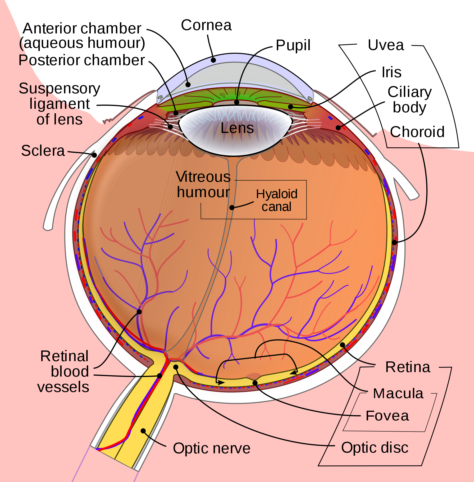
Test Characteristics of Point of Care Ultrasound for the Diagnosis of Retinal Detachment in the Emergency Department

Background

Retinal detachment (RD) is the final diagnosis for 3-4% of patients presenting to the Emergency Department (ED) with ocular complaints. Presenting symptoms most commonly include acute onset flashes and floaters, however, this presentation is not unique. The timely diagnosis and differentiation of RD from more common, benign, and similarly presenting processes, such as posterior vitreous detachment, is important in order to treat RD and prevent the sequela of permanent vision loss.

Point of care ultrasound (POCUS) has been successfully employed in the diagnosis of retinal pathology with high degrees of success according to observed test characteristics (sensitivity 97%-100%; specificity 83-100%) in emergency medicine (EM) literature. The generalizability of this data is limited, however, due to study features, including the use of highly experienced sonographers, inconsistent scanning protocols, and poor reference standards. This investigation seeks to derive the test characteristics for POCUS in the diagnosis of RD when used by a heterogeneous population of emergency physicians (EPs).

Test Characteristics of Point of Care Ultrasound for the Diagnosis of RetinalDetachment in the Emergency Department

Clinical Question

What are the test characteristics (sensitivity and specificity) of POCUS for the diagnosis of RD in patients presenting with chief complaint of flashes or floaters, when performed by a group of emergency physicians with varying degrees of ultrasound experience?

Methods & Study Design

  • Design
    • Prospective study using a convenience sample of patients presenting to the ED with a chief complaint of flashes or floaters in visual fields
  • Population
    • Conducted at Vancouver General Hospital, an urban academic tertiary care center
  • Inclusion Criteria
    • Patients presenting with chief complaint of acute (7 days or less) onset flashes or floaters in one or both eyes between March 2015 and September 2016
  • Exclusion Criteria
    • Age younger than 19 years, known diagnosis of RD, exam compromised due to advanced cataract in the affected eye, ophthalmologic surgery on affected eye within prior two weeks
  • Intervention
    • EP performed ocular POCUS with high-frequency linear transducer
    • Scan performed in both transverse and longitudinal plane with dynamic assessment of posterior chamber (patient looking left/right and up/down)
    • Positive or negative interpretation for RD was recorded
    • Reference Standard
      • Patients were referred to an ophthalmology resident who performed non-blinded assessment including a complete dilated retinal exam
      • Patients were then seen by a retina specialist blinded to the ED POCUS within 1 week, or for patients with a retinal tear or RD diagnosis, within 1 day
    • Standardized training session for emergency providers
      • EM attendings (20), fellows (2), and residents (8) of varying ultrasound experience received a 1 hour lecture on the use of POCUS to detect RD
      • All participating EPs performed one practice scan on a healthy volunteers
  • Outcomes
    • Primary outcome: Accuracy of the EP diagnosis with respect to the reference standard, the retina specialist diagnosis
    • Test characteristics: sensitivity, specificity, diagnostic accuracy, LR+, and LR- 

Results

Flow of Patients Through Study
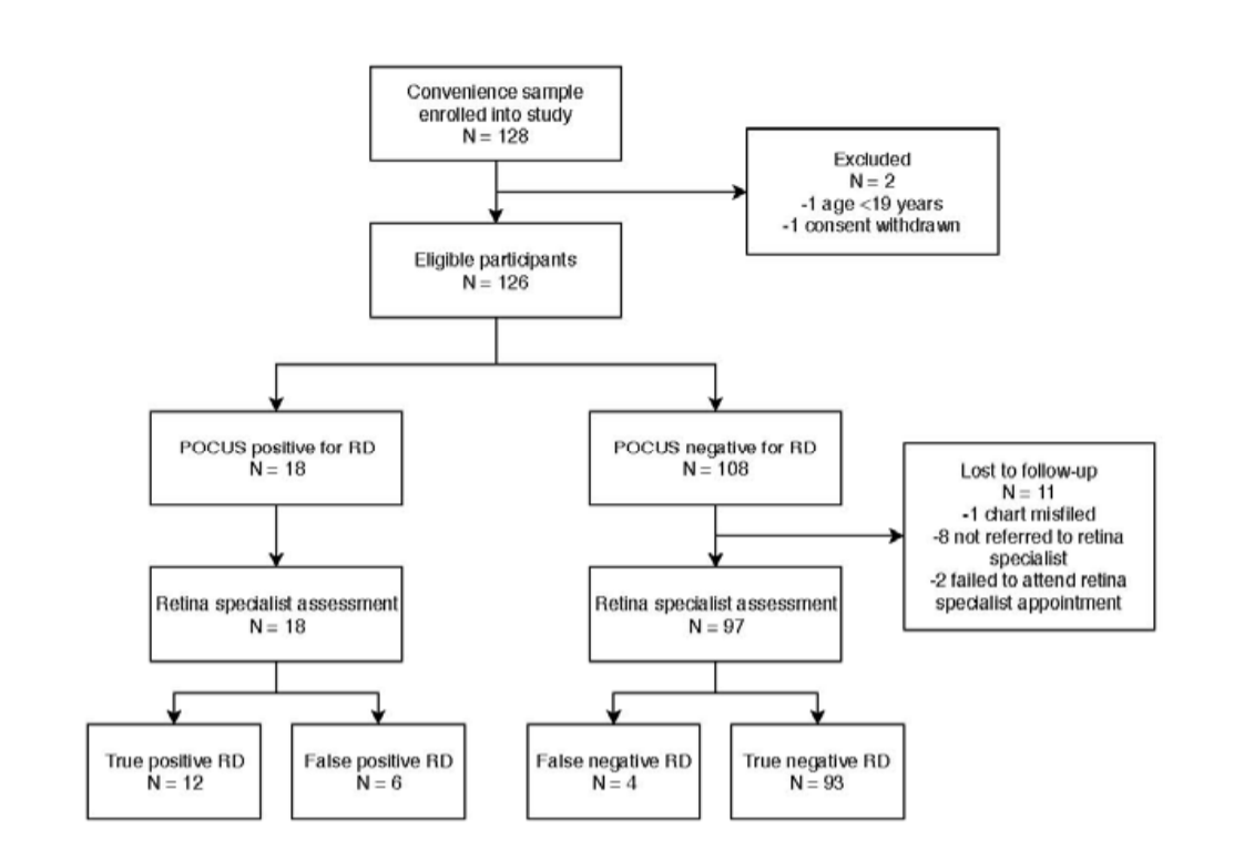
Primary analysis

    • Sensitivity: 75% (95% CI 48-93%)
    • Specificity: 94% (95% CI 87-98%)
    • Diagnostic accuracy: 91% (95% CI 85-96%)
    • LR-positive: 12.4 (95% CI 5.4-28.3)
    • LR-negative: 0.27 (95% CI 0.11-0.62)

Secondary analyses

    • Test characteristics by level of training
      • Residents and fellows: 100% sensitivity, 95% specificity
      • Attending physicians: 71% sensitivity, 94% specificity
    • Test characteristics by number of patients enrolled by EP
      • 1-2 patients enrolled: 80% sensitivity, 71% specificity
      • 3 or more patients enrolled: 73% sensitivity, 98% specificity

Limitations

    • Insufficiently powered for the secondary analyses
    • Single program study limits generalizability
    • Prior ultrasound experience was not explicitly assessed
    • RD is not always classically presenting, starting with a population defined by classic symptoms may influence observed test characteristics

Authors Conclusion

“In a heterogeneous group of EPs with varying ultrasound experience, POCUS demonstrates high specificity but only intermediate sensitivity for the detection of RD. A negative POCUS scan in the ED performed by a heterogeneous group of providers after a one-hour POCUS didactic is not sufficiently sensitive to rule out RD in a patient with new onset flashes or floaters.”

Our Conclusion

This study demonstrates that emergency physicians of varying training levels and ultrasound experience can successfully employ POCUS in the diagnosis of RD after only a short training session. By incorporating POCUS into the workup of patients presenting with ocular complaints characteristic of RD, true pathology can be identified with high specificity. Appropriate care can then be mobilized expeditiously in these scan-positive patients in order to prevent the permanent vision loss associated with this condition.

Indeed, a 74% sensitivity is too low for POCUS to reliably be utilized by a heterogeneous population of EPs as a tool to rule-out RD, especially given the consequences of a missed diagnosis. It would be reasonable practice, therefore, as the authors suggest, for all patients with new onset flashes and/or floaters to continue be referred for further ophthalmologic evaluation to definitively rule-out RD and other conditions at-risk for progression to RD. It should also be noted, however, that a trend towards increased specificity was observed amongst physicians who enrolled more patients in this study. Taken in context with test characteristics reported in prior literature, these findings may suggest that specificity can be improved upon with experience, and in the hands of a trained sonographer, POCUS may also be used as a tool to reliably rule-out RD.

The Bottom Line

Emergency providers can reliably use point-of-care ultrasound to diagnose retinal detachment with high specificity after a short, one-time training course, but must recognize the limitations of POCUS as a tool to rule-out RD in this setting, given a relatively low sensitivity when used for this purpose.

Authors

This post was written by Oretunlewa Soyinka, MS4 at UCSD. Review and further commentary was provided by Cameron Smyres, MD, Ultrasound Fellow at UCSD.

References

1 .  Hikichi T, Hirokawa H, Kado M, et al. Comparison of the prevalence of posterior vitreous
detachment in whites and Japanese. Ophthalmic Surg 1995; 26:39-43.

2.  Hollands H, Johnson D, Brox AC, et al. Acute-onset floaters and flashes: is this patient at
risk for retinal detachment? JAMA 2009; 302:2243-9

3.  Alotaibi AG, Osman EA, Allam KH, et al. One month outcome of ocular related
emergencies in a tertiary hospital in Central Saudi Arabia. Saudi Med J 2011; 32:1256-60.

4.  Mitry D, Charteris DG, Fleck BW, et al. The epidemiology of rhegmatogenous retinal
detachment: geographical variation and clinical associations. Br J Ophthalmol 2010;
94:678-84.

The Predictive Value of Bedside Ultrasound to Restore Spontaneous Circulation in Patients with PEA: A Systematic Review and Meta-Analysis

Background

Cardiac arrest remains one of the leading causes of death in the United States and is frequently encountered in the emergency department (ED). It is defined as cessation of cardiac function and lack of circulation. Cardiopulmonary resuscitation (CPR) improves outcomes especially if it is performed within minutes of cardiac arrest. According to recent American Heart Association (AHA) statistics, approximately,  10.6% of patients who experience cardiac arrest survive to hospital discharge [1]. On the other hand, pulseless electrical activity (PEA) is a form of cardiac arrest in which patients continue to have organized cardiac electrical activity without a palpable pulse. This patient population's overall survival is much lower with 2.4% of patients surviving to hospital discharge [2]. Until recently, there has been an incomplete understanding of the the term PEA and what this means physiologically. With the advent of ultrasound (US), there has now been elucidation of two forms of PEA. True-PEA (tPEA) which lacks cardiac activity on US, has poor survival rates, while pseudo-PEA (pPEA) which demonstrates some cardiac activity on US,  shows improved survival,  potentially due to altering standard ACLS protocol driven management. The following study specifically looks at the data evaluating the predictive value of US in patients presenting in cardiac arrest with PEA.

The predictive value of bedside ultrasound to restore spontaneous circulation in patients with pulseless electrical activity: A systematic review and meta-analysis.

Clinical Question

Does bedside US predict the restoration of spontaneous circulation in patients with pulseless electrical activity?

Methods & Study Design

  • Design
    • Systematic review and meta-analysis
    • Data from MEDLINE, EMBASE, Cochrane library databases (inception to June 2017)
    • Statistical analysis
      • Review Manager 5.4 and Stata 12
      • I2 statistics to assess heterogeneity
      • Random effects model for all polled outcome measures
      • Begg’s test for publication bias
  • Study Eligibility Criteria
    • Adults with PEA
    • Cardiac US was used to detect cardiac activity
    • ROSC defined as primary outcome
    • Prospective/ observational studies
    • Written in English
    • 2x2 contingency table can be formed from data

Results

Included Studies

    • 11 studies with 777 patients with PEA included
    • 230 patients had ROSC
    • 42/343 "true-PEA" patients had ROSC
    • 188/434 "pseudo-PEA" patients had ROSC
    • Patients with pPEA were 4.35x more likely to experience ROSC than those tPEA (Risk ratio 4.35, confidence interval 2.20-8.63, p<0.00001, significant statistical heterogeneity I2= 60%)

Limitations

    • Significant heterogeneity amongst the 11 studies
      • 4 studies enrolled both trauma and non-trauma patients
      • In 3 studies, US evaluation occurred in the pre-hospital setting
    • Large confidence interval
    • Small pooled sample size
    • Varying protocols and US views used in different studies to determine cardiac activity
    • Varying definition of ROSC between studies

Authors Conclusion


"In cardiac arrest patients who present with PEA, bedside US has an important value in predicting ROSC. The presence of cardiac activity in PEA patients may encourage more aggressive resuscitation. Alternatively, the absence of cardiac activity under US could be promoted as a way of confirming a poor prognosis and used to support the decision to terminate resuscitative efforts."

Our Conclusion

This study found that patients in cardiac arrest with pPEA (i.e. cardiac motion on ultrasound) have higher ROSC than those with tPEA (i.e. no cardiac motion on ultrasound). The exact risk ratio for ROSC quoted in their results should be interpreted with caution since this meta-analysis included studies with vastly different characteristics. The 11 studies included took place in 9 different countries over the span of 15 years, included different US views (subxiphoid, parasternal), varied settings (pre-hospital and in-hospital US studies), varied patient populations (some studies included traumatic cardiac arrest) and had varying US operator experience. Additionally, other factors such as time to initiation of CPR, length of CPR, and the previous health of the patient were not accounted for. These limitations can affect the accuracy of the risk ratio presented in this study.  That being said, even with significant heterogeneity in this study, resulting in a very wide confidence interval, the lower limit of the risk ratio (2.20) still finds statistical significance for higher rate of ROSC in patients with pPEA compared to patients with tPEA.

This study essentially confirms what is already known from previous data (specifically the Gaspari study which represents the majority of patients in this meta-analysis) but fails to address the big question of "Does US guided resuscitation provide a mortality benefit in the management of cardiac arrest?" This is a complex question that takes into account multiple other questions including the debate over US increasing interruptions in chest compressions, the use of US to identify immediately reversible causes of cardiac arrest (i.e. tamponade, massive PE) , the true definition of cardiac standstill (which calls the results of all cardiac arrest studies thus far into questions), and ultimately, can US be used to determine if further resuscitation is futile? As with all advances in technology, finding the right niche to benefit the patient is of upmost importance and at this point in time, the utility of US in cardiac arrest remains to be determined.

The Bottom Line

Bedside ultrasound can be used to determine pPEA from tPEA in patients with cardiac arrest. This may help guide resuscitation efforts as patients with pPEA have increased rates of ROSC.

Authors

This post was written by Tina Vajdi, MS4 at UCSD. Review and further commentary was provided by Michael Macias, MD, Ultrasound Fellow at UCSD.

References

    1. Mozaffarian D, Benjamin EJ, Go AS, Arnett DK, Blaha MJ, Cushman M et al. Heart Disease and Stroke Statistics-2016 Update: A Report From the American Heart Association. Circulation 2016; 133(4):e38– e360. https://doi.org/10.1161/CIR.0000000000000350 PMID: 26673558
    2. Engdahl J, Bang A, Lindqvist J, Herlitz J. Factors affecting short- and long-term prognosis among 1069 patients with out-of-hospital cardiac arrest and pulseless electrical activity. Resuscitation 2001; 51 (1):17–25. PMID: 11719169
    3. Gaspari R, e. (2018). Emergency department point-of-care ultrasound in out-of-hospital and in-ED cardiac arrest. - PubMed - NCBI . Ncbi.nlm.nih.gov. Retrieved 20 April 2018, from https://www.ncbi.nlm.nih.gov/pubmed/27693280

Can The Degree of Hydronephrosis on Ultrasound Predict Kidney Stone Size?

Background

Symptomatic renal colic is a common complaint presenting to the emergency department (ED), with a rate of 126 to 226 per 100,000 ED visits [1]. In the ED, CT is frequently used to make the definitive diagnosis as it allows for determination of stone size and location, degree of hydronephrosis, and evaluation of other pathology that may mimic renal colic. However this is a particularly worrisome approach in patients with recurrent ureteral stones who have been exposed to numerous previous CT imaging studies. Previous data has shown that emergency physician performed ultrasound is accurate at identifying hydronephrosis, which in combination with hematuria, is sufficient for the diagnosis of renal colic [2,3]. Furthermore,  an ultrasound first approach has been shown to be safe and reasonable as an initial evaluation for suspected renal colic [4]. What ultrasound does not tell us about renal colic is the size of the ureteral stone, which can be useful in determining the need for immediate intervention versus medical management. The following study seeks to determine if the degree of hydronephrosis seen on ultrasound performed by emergency physicians, can be predictive of ureteral stone size. 

Can the degree of hydronephrosis on ultrasound predict kidney stone size?

Clinical Question

Can the degree of hydronephrosis on ultrasound predict kidney stone size?

Methods & Study Design

  • Design
    • Retrospective chart review of emergency department (ED) patients at a single academic medical center
  • Population + Inclusion Criteria 
    • Adult patient presenting to the emergency department who had confirmed ureterolithiasis on noncontrast CT and a focused emergency renal ultrasound performed
  • Exclusion criteria
    • No specific criteria
  • Intervention
    • A focused renal ultrasound was performed in the ED by an emergency medicine resident or attending to evaluate for the presence of hydronephrosis as an indicator of obstructive ureterolithiasis
    • All ultrasound examinations were subsequently reviewed for quality assurance by an emergency ultrasound fellowship trained emergency physician
  • Outcomes
    • Each focused renal ultrasound classified the degree of hydronephrosis as none, mild, moderate, or severe and this was compared to the ureteral stone size on noncontrast CT
      • Definitions:
        • Mild hydronephrosis was defined as enlargement of the calices withpreservation of the renal papillae
        • Moderate hydronephrosis was defined as rounding of the calices with obliteration of therenal papillae
        • Severe hydronephrosis was defined as caliceal ballooning with cortical thinning
    • Ureteral stone size was stratified into 2 groups, those 5mm or smaller and those larger than 5 mm, based on the likelihood of successfully spontaneous stone passage

Results

Increasing degree of hydronephrosis seen on focused ultrasound was associated with an increasing proportion of ureteral calculi larger than 5 mm. 113 (87.6%) patients with less severe hydronephrosis  (none or mild) had ureteral calculi 5 mm or smaller. Of the remaining 16 (12.4%) patients with less severe hydronephrosis, none of these patients had ureteral stones larger than 10 mm. There was good interobserver agreement between the degree of hydronephrosis as determined by the performing emergency physician and the quality assurance review (k = 0.847).

Strengths & Limitations

  • Strengths
    • Majority of ultrasound examinations performed by ED physicians making this applicable to point-of-care ultrasound
    • Gold standard was size of ureteral stone on noncontrast CT
    • Good interobserver agreement between ED ultrasound operator and quality assurance review
  • Limitations
    • Retrospective chart review
    • This study only enrolled patients who both a focused renal ultrasound and confirmed ureterolithiasis on noncontrast CT; this would have missed patients who only had either a focused renal ultrasound or noncontrast CT alone (selection bias)
    • No patient centered outcomes data

Authors Conclusion

"In conclusion, our results demonstrate a relationship between the degree of hydronephrosis as determined by emergency physicians on focused emergency ultrasound and ureteral calculi size; patients with less severe hydronephrosis were less likely to have larger ureteral calculi. This suggests that ultrasound can help identify many, but not all, patients who are at lower risk for having larger ureteral calculi.

Our Conclusion

This paper identifies a correlation between the degree of hydronephrosis on ultrasound and ureteral stone size seen on noncontrast CT. Essentially, patients with minimal or no hydronephrosis are very unlikely to have have a large (>5 mm) ureteral stone. Unfortunately, focused ultrasound is not perfect, and in this study  ~12.4% of patients with minimal or no hydronephrosis still had a large ureteral stone. What I found reassuring was that in this group, none of the patients had a ureteral stone > 10 mm, which at most institutions is the cut off for allowing a trial of passage. Even dissecting the data further, of the patients with moderate hydronephrosis, only 2 out of 43 (4.6%) patients had a stone > 10 mm.

This study suggests that focused renal ultrasound can be used to screen patients with suspected renal colic and potentially avoid an unnecessary CT scan. As with any focused ultrasound, the decision to obtain a CT should not be based solely the degree of hydronephrosis but also in conjunction with the clinical history, physical exam and other pertinent factors (previous ureteral stone, previous need for stone intervention, other concerning diagnoses on differential, pain control, institutional culture, urinalysis, etc). 

The Bottom Line

Ultrasound can be used to identify many, but not all, patients who are at lower risk for having larger ureteral calculi. 

Authors

This post was written by Michael Macias, MD, Ultrasound Fellow at UCSD.

References

    1. Teichman JMH. Clinical practice. Acute renal colic from ureteral calculus. N Engl J Med 2004;350:684-93.

    2. Rosen CL, Brown DFM, Sagarin MJ, et al. Ultrasonography by emergency physicians in patients with suspected ureteral colic. J Emerg Med 1998;16:865-70.

    3. Gaspari RJ, Horst K. Emergency ultrasound and urinalysis in the evaluation of flank pain. Acad Emerg Med 2005;12:1180-4.

    4. Smith-Bindman R, e. (2018). Ultrasonography versus computed tomography for suspected nephrolithiasis. - PubMed - NCBI . Ncbi.nlm.nih.gov. Retrieved 3 March 2018, from https://www.ncbi.nlm.nih.gov/pubmed/25229916
    5. S, G. (2018). Can the degree of hydronephrosis on ultrasound predict kidney stone size? - PubMed - NCBI . Ncbi.nlm.nih.gov. Retrieved 3 March 2018, from https://www.ncbi.nlm.nih.gov/pubmed/20837260

Free Fluid in Morison’s Pouch on Bedside Ultrasound Predicts Need for Operative Intervention in Suspected Ectopic Pregnancy

Background

Vaginal bleeding and abdominal pain are common presenting symptoms to the emergency department (ED) in women in early pregnancy. While the majority of pregnant patients presenting with these symptoms are stable, a small subset of this group represents a ruptured ectopic pregnancy with a potential to rapidly decompensate, resulting in high rates of morbidity and mortality. Therefore it is critical for this diagnosis to be identified both accurately and rapidly.

The Focused Assessment of Sonography in Trauma (FAST) exam has been well studied in the acute trauma setting to identify internal bleeding however there is not much data on its use in the non trauma setting. In theory, it makes intuitive sense that it should perform similarly and be able to accurately identify significant non traumatic pelvic and intraperitoneal bleeding.

In a previous study, emergency physician (EP) performed bedside ultrasound (US) in suspected ectopic pregnancy was found to  decrease the time to both diagnosis and treatment [1]. The following study seeks to prospectively investigate the significance of positive fluid in Morison's pouch during transabdominal US examination performed by EPs in patients with suspected ectopic pregnancy. 

Free Fluid in Morison’s Pouch on Bedside Ultrasound Predicts Need for Operative Intervention in Suspected Ectopic Pregnancy‌

Clinical Question

Is EP performed US with identification of free fluid in Morison's pouch predictive of the need for operative intervention in suspected ectopic pregnancy?

Methods & Study Design

  • Design
    • Prospective observational study
  • Population
    • Conducted at Yale-New Haven Hospital, an urban Level 1 trauma center and teaching hospital
    • Pregnant women presenting to the ED in whom there was a suspicion of an ectopic pregnancy
  • Inclusion criteria
    • Female patients with positive pregnancy test who presented in first trimester with abdominal pain and/or vaginal bleeding and requiring further imaging or consultation
  • Exclusion criteria
    • No specific criteria
  • Intervention
    • EP performed transabdominal and transpelvic US evaluation for:
      • Free fluid in Morison's pouch (positive, negative or indeterminate)
      • Presence of intrauterine pregnancy (IUP), or no definitive IUP
      • Free fluid in the cul-de-sac (present, absent)
  • Outcomes
    • Follow up and chart review was performed by independent study investigators, blinded to ED US results, ultimately classifying the final outcome as ectopic or non ectopic pregnancy and further defining the management as operative or medical.

Results

Patient Flow Diagram 

Ultrasound Findings and Clinical Characteristics  of Patients


Strengths & Limitations

  • Strengths
    • Performed in ED based population
    • All ultrasound examinations performed by ED physicians making this applicable to point-of-care ultrasound
  • Limitations
    • Potential selection bias given that rate of ectopic pregnancy in study population was higher than most published rates
    • Not truly observational study as treating physicians were not blinded to ED US results
    • Some patients lost to follow up

Authors Conclusion

"Free intraperitoneal fluid found in Morison’s pouch in patients with suspected ectopic pregnancy may be rapidly identified at the bedside by an EP-performed US and predicts the need for operative intervention. Transabdominal pelvic US may show an IUP in more than one third of patients with suspected ectopic pregnancy."

Our Conclusion

While this paper does not have the methodological prowess of a multicenter randomized control trial, it accurately answers an important question with respect to positive fluid in Morison's pouch on ED US and the need for operative intervention in ectopic pregnancy. This study also highlights the niche of point-of-care ultrasound and why it is so critical to our practice as emergency medicine providers. Unlike radiological studies, which require increased time and may be difficult to obtain in critical patients, point-of-care ultrasound allows rapid identification of key findings that allow for early diagnosis and decision making.

Based on this study, the addition of positive fluid seen in Morison's pouch during ED US for suspected ectopic pregnancy now adds on a strong predictor for the need for operative intervention. This is especially important in unstable and hypotensive patients.

However, there are several caveats to understand when interpreting this data. First, all the patients in this study were enrolled after having a positive pregnancy test, therefore, prior to assuming that a young female with free fluid in her abdomen is from a ruptured ectopic pregnancy, a pregnancy must first be confirmed.  Second, not all free fluid in Morison's pouch in a pregnant woman is due to an ectopic pregnancy. In this data set, there was one patient that had a definitive IUP and free fluid in Morison's pouch which was found to be from a ruptured corpus luteal cyst. There are also case reports of splenic artery aneurysm rupture in pregnancy that could mimic a ruptured ectopic [2]. If an IUP is identified on transabdominal ultrasound, unless the patient is receiving advanced reproductive techniques, alternative explanations for the free fluid should at least be considered before a heterotopic pregnancy is presumed. Third, while this study demonstrates excellent specificity of positive fluid in Morison's pouch and the need for operative intervention in suspected ectopic pregnancy, it has very poor sensitivity. Therefore, ED US should be used as a rule in technique and does not replace formal ultrasound and obstetrical consultation if the diagnosis is not clear.

The Bottom Line

Free fluid found in Morison’s pouch in patients with suspected ectopic pregnancy can be rapidly identified at the bedside by emergency providers and predicts the need for operative intervention.


Authors

This post was written by Michael Macias, MD, Ultrasound Fellow at UCSD.

References

    1. Rodgerson JD, Heegaard WG, Plummer D, Hicks J, Clinton J, Sterner S. Emergency department right upper quadrant ultrasound is associated with a reduced time to diagnosis and treatment of ruptured ectopic pregnancies. Acad Emerg Med. 2001; 8:331–6.
    2. Lynch MJ, Woodford NW. Rupture of a splenic artery aneurysm during pregnancy with maternal and foetal death: a case report. Med Sci Law. 2008;48:(4)342-5.
    3. Moore C et al. Free fluid in Morison's pouch on bedside ultrasound predicts need for operative intervention in suspected ectopic pregnancy. Acad Emerg Med. 2007; 8: 755-8

Case # 13: What Lies Beneath

A 30 year old male presents to the emergency department after blunt trauma to the face from an altercation. He notes he was punched several times in the face but did not pass out. His exam is notable for significant right periorbital ecchymosis and edema with inability to open his eye. You are unable to perform a direct eye exam given the significant periorbital swelling.  A CT maxillofacial is performed which shows an isolated right inferior orbital wall fracture.

Vitals: T 98.6 HR 85 BP 142/81  RR 14 O2 98% on RA

Prior to ENT consultation, a bedside ultrasound of the orbits is performed.  In spite of being unable to open the eye, what can you tell your consultant regarding your exam?

ezgif.com-resize

ezgif.com-resize (1)

Answer and Learning Points

Answer

Using ultrasound as an adjunct to your exam, you are able to tell the consultant that there is a normal appearing, reactive pupil and that the extra-ocular movements of the eye are intact. The consultant is appreciative over the phone and is happy to come in and see the patient whom after evaluation is discharged home with close outpatient follow up.

Learning Points

It is often the case where a patient suffers such significant facial trauma that a complete physical exam of the orbit due to periorbital swelling is not possible. Ultrasound can be a critical tool in these cases to provide useful information to assess for multiple potential pathologies. Previous studies have shown the ability of ocular ultrasound in trauma to detect elevated intracranial pressure (via optic nerve sheath diameter), retinal detachment, vitreous hemorrhage, and retrobulbar hematoma. It can also be used for early detection of muscular entrapment in the case of an orbital wall fracture, as well as performed serially for pupillary response in patients with significant neurological injury at risk for deterioration and potential herniation.

  • To evaluate extraocular movements:
    • Prepare the patient by laying the bed backwards and having their face parallel to the ceiling,  supporting the patient's head and neck with a pillow or blanket.
    • (Optional) Place a tegaderm over the eye. If you do, ensure there is no air between the tegaderm and the eyelid.
    • Place a small amount of ultrasound gel on the closed eyelid  and prepare the linear probe with the gain turned almost all the way up.
    • Stabilize your hand on the patient's nasal bridge or zygoma, with the probe marker to your left, and place the probe transverse on the orbit with minimal pressure being applied directly to the eye. This is very important in trauma as the area is likely painful and theoretically the patient could have a ruptured globe.
    • Adjust the depth to ensure the optic nerve is just visualized at the bottom of the screen. The anterior chamber and lens should be used as visual landmarks to ensure you are in proper location.
    • Next, have the patient look left and right, then turn the probe to a sagittal orientation and have the patient look up and down. During these maneuvers you should be evaluating for symmetric movements of the orbit in each direction.
    • If you do not appreciate symmetric movements of the orbit in all directions then you may have entrapment of an extraocular muscle.
  • To evaluate for pupillary response and shape:
    • Be sure to dim the lights in the room prior to performing this exam to allow for an adequate pupillary response.
    • Gently apply the linear probe with gel in a transverse plane just inferior to the eye, angling superiorly towards the patient's head (Depending on the location of the swelling around the eye, you can also place the probe superior to the eye, angling inferiorly towards the patient's feet).
    • Keep flattening out your probe angle relative to the skin until you have a cross section of the pupil and iris in view.
    • The pupil should be evaluated for symmetry as an asymmetric or oblong pupil could suggest globe rupture. You can then shine a light in the affected or non-affected eye (consensual light reflex) and observe the pupil for constriction.


Author

This post was written by Michael Macias, MD, Ultrasound Fellow at UCSD.

References

    1. Blaivas M. Bedside emergency department ultrasonography in the evaluation of ocular pathology. Acad Emerg Med 2000;7:947-50.
    2. Blaivas M, Theodoro D, Sierzenski P. A study of bedside ocular ultrasonography in the emergency department. Acad Emerg Med 2002;9(8):791-9.
    3. Kimberly HH, Shah S, Marill K, Noble V. Correlation of optic nerve sheath diameter with direct measurement of intracranial pressure. Acad Emerg Med 2008;15(2):201-4.
    4. Tayal VS, Neulander M, Norton HJ, et al. Emergency department sonographic measurement of optic nerve sheath diameter to detect findings of increased intracranial pressure in adult head injury patients. Ann Emerg Med 2007;49(4):508-14.
    5. Harries A, et al. Ultrasound assessment of extraocular movements and pupillary light reflex in ocular trauma. Am J Emerg Med 2010 28(8):956-9.

Prospective Outcomes of Pregnant ED Patients with Documented Fetal Cardiac Activity on Ultrasound

Background

Vaginal bleeding is a common cause of presentation to the emergency department (ED), and is the leading cause of presentations to the ED among patients with first trimester pregnancy [1]. It is reported that up to 25% of pregnancies have some bleeding within the first trimester [2]. Based on previous data, bleeding in the first trimester represents an increased risk of spontaneous abortion (SAB), with up to 50% of women losing the pregnancy [3]. However, patients with first trimester bleeding and a documented intrauterine pregnancy (IUP)  with fetal heart tones (FHTs), represent a subset of first trimester bleeding patients with improved outcomes, with rates of SAB ranging from 11.1-16.4% [4-5]. These previous studies were performed in outpatient obstetrical clinics and no prospective data exists with respect to outcomes in this subset of patients presenting to the emergency department. 

Prospective Outcomes of Pregnant ED Patients with Documented Fetal Cardiac Activity on US

Clinical Question

What is the rate of SAB in pregnant women presenting to the ED with first trimester bleeding and a documented IUP with cardiac activity?

Methods & Study Design

  • Design
    • Prospective observational study
    • Convenience sample of pregnant patients presenting to the University of Utah ED from January 1, 2008 through April 30, 2010.
  • Population
    • Pregnant women presenting to the ED with abdominal pain and/or bleeding
  • Inclusion criteria
    • Ultrasound (performed by ED physician at bedside or formal radiology study) demonstrating an IUP with FHTs and whose pregnancy dates placed them in the first trimester (< 13 weeks)
  • Exclusion criteria
    • No specific criteria
  • Intervention
    • Ultrasound demonstrating IUP with FHTs
  • Outcomes
    • Rate of SAB at 30 days after ED visit
    • Patients were contacted by telephone at least 30 days after their ED visit and asked about the status of their pregnancy

Results

Strengths & Limitations

  • Strengths
    • Performed in ED based population
    • Majority of ultrasound examinations performed by ED physicians making this applicable to point-of-care ultrasound
  • Limitations
    • Performed at single academic center
    • Low patient enrollment leading to large CI for rate of SAB
    • 85.9% patient follow up rate
    • Patients only followed out to 30 days after ED visit

Authors Conclusion

"In this prospective study of ED patients with first trimester bleeding and/or pain, we found that patients who had an IUP and FHTs by ED US had a 14.8% rate of SAB at 30 days. These findings may help to better define risk of SAB after first-trimester bleeding and allow us to provide more accurate counseling and prognostic information to pregnant ED patients presenting with these symptoms.”

Our Conclusion

This is an excellent paper that helps provide emergency medicine providers with prognostic information  regarding women presenting to the ED during first trimester pregnancy with vaginal bleeding and a documented IUP with FHTs. Often in emergency medicine we are focused on ruling out the life threatening diagnoses, in the above scenario, ectopic pregnancy, and it can be easy to lose sight of other important aspects of patient care. This paper helps refocus our attention and gives us important data to be able to provide an already anxious patient with some useful information on the potential expected course of their pregnancy. With this data, we are now able to better define the risk of SAB after first trimester bleeding and provide improved counseling and prognostic information to these patients.

The Bottom Line

In ED patients with first trimester bleeding, those that have an IUP and FHTs by ED ultrasound have ~15% rate of SAB at 30 days. 

Authors

This post was written by Michael Macias, MD, Ultrasound Fellow at UCSD.

References

    1. Dighe M, Cuevas C, Moshiri M, Dubinsky T, Dogra VS. Sonography in first trimester bleeding. J Clin Ultrasound 2008;36(6):352-66.
    2. Hasan R, Baird DD, Herring AH, Olshan AF, Jonsson Funk ML, Hartmann KE. Patterns and predictors of vaginal bleeding in the first trimester of pregnancy. Ann Epidemiol 2010;20(7):524-31.
    3. Dideriksen KL, Lidegaard O, Langhoff-Roos J. First trimester vaginal bleeding and complications later in pregnancy. Obstet Gynecol 2010;115(5):935-44.
    4. Poulose T, Richardson R, Ewings P, Fox R. Probability of early pregnancy loss in women with vaginal bleeding and a singleton live fetus at ultrasound scan. J Obstet Gynaecol 2006;26(8):782-4.
    5. Siddiqi TA, Caligaris JT, Miodovnik M, Holroyde JC, Mimouni F.Rate of spontaneous abortion after first trimester sonographic demonstration of fetal cardiac activity. Am J Perinatol 1988;5(1):1-4.
    6. Mallin M, e. (2018). Prospective outcomes of pregnant ED patients with documented fetal cardiac activity on ultrasound. - PubMed - NCBI . Ncbi.nlm.nih.gov. Retrieved 26 January 2018, from https://www.ncbi.nlm.nih.gov/pubmed/21334156

Case # 12: Bilateral Vision Loss

A 45 year old male with poorly controlled DM presents with bilateral vision loss. His right eye vision acutely worsened 3 days ago with the sensation of a curtain moving back and forth across his visual field. Today his left eye vision acutely worsened with flashes and floaters occurring. He denies any trauma, headache, or new medications.

Vitals: T 98.6 HR 90 BP 149/87  RR 16 O2 98% on RA

A bedside ultrasound of the orbits is performed,  what is the next best step in management?

Left Eye

Left Eye

Right Eye

Right Eye

Answer and Learning Points

Answer

The ultrasound clips demonstrate hypoechoic material in the orbits bilaterally, swirling around with subtle eye movement. This is consistent with bilateral vitreous hemorrhage. The diagnosis was discussed with the patient and he was referred to ophthalmology clinic for dilated eye exam in 24 hours.

Learning Points

Vitreous hemorrhage is a common diagnosis (though usually unilateral) seen in poorly controlled diabetes. The most frequent etiologies include proliferative diabetic retinopathy, posterior vitreous detachment, and ocular trauma, with trauma more common in patients under the age of 40. Since it is difficult to obtain  a good physical exam of the posterior aspects of the eye without a dilated exam, there is high utility in the use of point of care ultrasound in evaluating for acute pathology.  It can be used to distinguish vitreous hemorrhage and retinal detachment, which have significantly different prognoses and treatment pathways. To perform an ocular ultrasound, follow these steps:

    1. Prepare the patient by laying the bed backwards and having their face parallel to the ceiling,  supporting the patient's head and neck with a pillow or blanket.
    2. Place a tegaderm over the eye (optional). If you do, ensure there is no air between the tegaderm and the eyelid.
    3. Place the ultrasound gel on the tegaderm and prepare the linear probe with the gain turned almost all the way up (this will help you visualize both retinal detachment and vitreous hemorrhage.
    4. Stabilize your hand on the patient's nasal bridge or zygoma, with the probe marker to your left, and place the probe transverse on the orbit with minimal pressure being applied directly to the eye.
    5. Adjust the depth to ensure the optic nerve is just visualized at the bottom of the screen. The anterior chamber and lens should be used as visual landmarks to ensure you are in proper location. Next, have the patient look up, down , left and right (oculokinetic echography), to assess for any abnormalities in the posterior aspects of the eye.
    6. Repeat this technique with the probe marker pointed superiorly and have the patient again look in all directions.

Retinal detachment: The common POCUS findings include a thin linear structure tethered to the optic nerve.  It flaps back and forth as the eye is moved giving it the appearance of “swaying seaweed”. This is an ophthalmologic emergency, especially if the macula is still attached,  the ophthalmologist should be immediately consulted.

Vitreous hemorrhage: You will notice a diffuse mobile opacity often described as a “snow globe” that is exacerbated with moving the eye from side to side. If this is seen in a diabetic patient with floaters, there is a high likelihood that the diagnosis is a vitreous hemorrhage. These patients will still need follow up with ophthalmology for further management, but typically there will not be an emergent intervention.

Author

This post was written by Sam Frenkel, MD, PGY-2 UCSD EM. It was reviewed by Michael Macias, MD, Ultrasound Fellow at UCSD.

References

    1. Yoonessi R, Hussain A, Jang TB. Bedside ocular ultrasound for the detection of retinal detachment in the emergency department. Acad Emerg Med. 2010;17(9):913-7.
    2. Dawson, Mallin. Introduction to Bedside Ultrasound, Volume 2. 2013. Apple iBook.
    3. Kilker B, Holst J, Hoffmann B. Bedside ocular ultrasound in the emergency department. Eur J Emerg Med. 2014;21(4):246-253.
    4. Shinar Z, Chan L, Orlinsky M. Use of ocular ultrasound for the evaluation of retinal detachment. J Emerg Med. 2011;40(1):53-57. 

Ultrasound Guided Catheterization of the Radial Artery

Background

Arterial catheterization (the radial artery being the most common site)  is often performed in critically ill patients for hemodynamic monitoring and serial blood gas sampling, and is a core skill for critical care and emergency providers alike. While ultrasound guidance has become standard of care for  central venous catheterization, this is still not common practice for radial artery catheterization. In this critically ill patient population, there are often patient specific factors that make this procedure difficult using the palpation method, including hypotension, edema and obesity. Frequently, the palpation method requires multiple attempts which can result in arterial vasospasm, making further attempts even more difficult. This review article investigates whether their is a role for the addition of ultrasound guidance to radial artery catheterization. 

Ultrasound-Guided Catheterization of the Radial Artery 

Clinical Question

Does ultrasound guidance for radial artery catheterization improve first attempt success compared to the palpation method? 

Methods & Study Design

  • Design
    • Systematic Review and Meta-analysis of Randomized Controlled Trials
      • Article selection
        • Databases searched: EMBASE, CENTRAL, and Medline from inception through February 23, 2010
        • Critical care conference abstracts reviewed from 2005-2009
        • Experts in the field also contacted to seek additional articles
        • The methodologic quality of selected trials was appraised by two independent reviewers using the Jadad criteria
  • Population (See Figure 1)
  •  Heterogenous population of adults and pediatric patients in different clinical settings
  • Inclusion criteria
    • Randomized control trials comparing 2-D ultrasound guidance technique to traditional palpation technique for radial artery catheterization
  • Exclusion criteria
    • Trials evaluating use of doppler ultrasonography, marking techniques, or catheterization of arteries other than the radial artery were excluded
  • Intervention
    • Ultrasound guided radial artery catheterization
  • Outcomes
      • First-attempt success for radial artery catheterization

Results

    • A total of 4 RCTs were included in the final meta-analysis. Individual characteristics can be viewed in figure 2.
    • Pooled Findings
      • The pooled relative risk for ultrasound guided techniques was 1.71 (95% CI, 1.25-2.32). The forest plot can be seen in figure 3 with data provided in figure 4.

Strengths & Limitations

  • Strengths
    • Comprehensive literature search
    • Included only RCTs comparing traditional palpation technique to ultrasound guided technique for radial artery catheterization
    • Adequate sample size obtained which reached statistical significance with respect to outcome
    • Studies independently reviewed by two reviewers for inclusion in meta-analysis
  • Limitations
    • The patient populations were very heterogenous among RCTs (1 adult surgery population, 1 infant neurosurgery population, 1 adult ED population, 1 pediatric surgery population)
    • The operator populations were very heterogeneous among RCTs (anesthesia attendings/residents, emergency physicians, pediatric trainee and consultant anasthesiologists)
    • Lack of blinding

Author's Conclusions

 "Our meta-analysis clearly demonstrated a 71% increase in the likelihood of first-attempt success when using ultrasound guidance for radial artery catheterization."

Our Conclusions

Radial artery catheterization is fraught with error and barriers to success. Often this procedure will be performed in sick patients, with the operator encountering obesity, edema, and shock, all of which can contribute to difficulty palpating the radial pulse. Furthermore, even if the pulse is palpated, this can be unreliable at predicting underlying anatomy. Ultrasound allows direct visualization of the radial artery, including depth, diameter and surrounding structures, and allows for ongoing needle guidance. While not studied, in my personal experience I have also noted numerous occasions where the only sign of successful radial artery catheterization was direct visualization of the catheter tip in the radial artery (i.e. no blood seen in flash chamber) on ultrasound. If the standard palpation method was used, this would lead to a failed attempt.

It has become clear in many other instances that ultrasound adds safety and success to procedures routinely performed in the emergency department including: central venous access, thoracentesis, paracentesis, peripheral nerve blocks and arthrocentesis (of specific joints). This study allows us to confidently add radial artery catheterization to the list. While the patient population and operators in the study discussed are heterogeneous, it is reasonable to assume that emergency medicine providers who have experience with ultrasound guided procedures, will perform just as well, if not better than the study findings.  This generalization assumes ultrasound guided procedure experience and those without this experience may not show a benefit over the traditional palpation method for radial artery catheterization. This study does not mean that the palpation method is obsolete, rather, it suggests that ultrasound is a useful adjunct and likely adds success in patients with risk factors for difficult radial artery catheterization such as obesity, hypotension, edema or a difficult to palpate pulse.

The Bottom Line

Ultrasound guidance for radial artery catheterization shows a higher first-attempt success rate compared to the standard palpation method and should be considered by operators with other procedural ultrasound guidance experience. 

Authors

This post was written by Michael Macias, MD, Ultrasound Fellow at UCSD.

References

    1. Shiloh AL, e. (2018). Ultrasound-guided catheterization of the radial artery: a systematic review and meta-analysis of randomized controlled trials. - PubMed - NCBI Ncbi.nlm.nih.gov. Retrieved 13 January 2018, from https://www.ncbi.nlm.nih.gov/pubmed/20724734

Case # 11: Look and You Will Find

A 32 year old healthy female with no past medical history presents to the emergency department with left sided flank pain x 2 days.

Vitals: T 98.6 HR 72 BP 126/82  RR 12 O2 98% on RA

A bedside ultrasound of the left kidney is performed, what is the next best step in management?

Q40_Simple cyst

Answer and Learning Points

Answer

The ultrasound image demonstrates a simple cyst located in the cortex of the kidney. The cyst can be described as anechoic, homogenous, with thin and smooth walls, and would be a type I lesion according to the Bosniak classification system (image below). There is no evidence to suggest obstructing hydronephrosis. The Bosniak classification for renal cysts was developed in the 1980s as an attempt to standardize the description and management of complex renal lesions. Based on classification of the renal lesion, the likelihood of malignancy can also be predicted. While the Bosniak classification was initially described and validated with CT imaging, newer data suggests that ultrasound may be sufficient to follow renal cysts that are minimally complex (Bosniak I & II).

Learning Points


    • Given the bedside ultrasound demonstrates a Bosniak I lesion in the left kidney, the patient can be reassured that this finding is very unlikely to be malignant and she can be referred to a primary care provider for follow up in several weeks for formal outpatient renal ultrasound.
    • Incidental findings are frequently found on point of care ultrasound and while most of them are benign it is of utmost importance to ensure proper follow up when identified. Specifically with renal cysts, this is a common occurrence and most can be followed with renal ultrasound as long as they are simple (Bosniak I or II).
    • One pitfall to be aware of is that renal cysts can be mistaken for hydronephrosis and lead to unnecessary imaging and work up (especially in patients presenting with acute flank pain). Therefore it is critical to note the differences between a simple renal cyst and hydronephrosis. As seen in the comparison above renal cysts tend to (but not always) be located in the renal cortex and are both spherical and very well circumscribed. On the other hand, hydronephrosis is centrally located, and tends to branch outwards like a tree. If there is uncertainty, I recommend performing evaluation in both transverse and longitudinal planes to fully characterize your finding. The opposite kidney in the patient can also be used for comparison.

Author

This post was written by Michael Macias, MD, Ultrasound Fellow at UCSD.

References

1. Muglia VF, Westphalen AC. (2014) Bosniak classification for complex renal cysts: history and critical analysis. Radiol Bras 47(6): 368–373.

2. McGuire BB, Fitzpatrick JM. (2010) The diagnosis and management of complex renal cysts. Curr Opin Urol 20:349–354.

3. Case courtesy of Dr Matt Skalski, Radiopaedia.org. From the case rID: 20989

How To: DIY Ultrasound Guided Peripheral IV Phantom

Intro_Procedural_Ultrasound-06

Introduction

Ultrasound-guided venous access has become a core skill for both emergency medicine providers, and nursing staff, to help facilitate difficult access. While clinical experience is critical to learning this technique, phantoms are also a necessary component, specifically with regards to developing both fine-motor skills and spatial orientation. In this post, we provide a step-by-step guide to creating a high-quality, affordable, phantom that can be used for educational purposes at large conferences or for small group training sessions.

IMG_0267

1. The Set Up

The Mold: water, metamucil, gelatin, citric acid, blue food coloring

The Veins: animal balloons, red food coloring

The Container: supply case ( 5.08 x 5.25 x 1.9 )

The Tools: sharpie, scissors, syringe, sharp knife or razor blade, large whisk (not shown)

IMG_0279

2. Create your Container

We chose the above container because it only requires 500 ml of gelatin mixture, it is plastic (which allows us to cut into it), and it has a lid which allows for easy transport and keeps the phantom fresh.

What you will be doing is creating small slits in the sides of the container to allow for the vessels to "float" and avoid having to pour gelatin in two steps which is both time consuming and messy.

IMG_0270

 2a. Measure out vessel depth and location

I like to place the vessels 1.5 cm below the surface of the gelatin mold. Using the thick plastic line near the opening of the box, measure 1.5 cm down from here. Measure 4 cm from each side of the box and connect everything with lines. The areas that cross will be where your vessels will go. Repeat this process on opposite side.

  • IMG_0272
  • IMG_0273

2b. Puncture four small slits in container

Now it's time to make small cuts in the plastic container which will allow the vessels to "float." I sit by the stove when doing this and turn on vent above to prevent significant inhalation of burning plastic fumes. Hold the tip of sharp knife or razor knife directly in flame until it turns red. Then quickly push knife into plastic at each line intersection, making approximately a 7-8 mm slit. You may have to re-heat knife between punctures.

Your container is now ready to be used, onto creation of your vessels.


  • IMG_0274
  • IMG_0275

2. Create your Vessels

The red food coloring adds an extra element to your phantom by making the "flash" in the angio-catheter chamber visible.

Fill your syringe with colored water and inject into balloon until it is plump. Next, make sure to get all the air out of the balloon that may have become trapped by holding the balloon vertical, allowing the air to rise to the top, and then tying this off. Continue to tie off the ends of the balloon and trim down with scissors until the vessel is just longer than the width of your container.

Make sure to leave around 1 cm of balloon on each side of your knots as you will need this to pull the vessels through the slits in the container.

  • IMG_0277
  • IMG_0278
  • IMG_0279

3. Cannulate your Container 

Take each balloon vessel and thread the ends through the slits in your container. Note as shown in the image that the knot will get caught up on the slit. I use the end of my scissors to pry open the slit slightly, allowing the knot to be pulled through with a little bit of force.

Once the knot is through, pull the balloon vessel out about 1 cm. This will allow the container to "auto-seal." so you do not have to worry about leaking when pouring in the gelatin mold.

This is the hardest step in constructing your phantom so do not be dissuaded if it takes you a few tries.

IMG_0282

4. Make your Gelatin Mold

Now it's time to make your gelatin mold. Here are the amounts required for a 500 ml gelatin mold. You can multiply these amounts as needed if making multiple models at a time:

Water: 500 ml

Gelatin: 40 grams (~3.5 Tbsp)

Metamucil: 20 grams (~1.75 Tbsp)

Citric Acid: 1 Tbsp

Blue Food Coloring: 1 tsp

ezgif.com-optimize (1)

4a.  Combine ingredients

Heat up the water in a large pot until it just begins to boil. At this point turn down heat so that water stays warm but is not bubbling. Too much heat will cause the mixture to froth and overflow.

Now add your ingredients, I do the gelatin first as this is the most difficult.  It is critical to whisk the water the entire time while adding the gelatin slowly. If you dump the gelatin in all at once, it will form large clumps and ruin your phantom. I would also follow this same technique when adding the other ingredients.

Once all ingredients have been well combined and your mixture has thickened you are ready to pour your gelatin mold into your container.

  • the pour
  • IMG_0289

4b. Pour the mold

Pour gelatin mold into your container, up to the thick plastic line. Allow the mold to cool for 10-15 minutes, then close lid and place in the fridge overnight to set.

The next day your mold will be ready to use!

  • ezgif.com-optimize (2)
  • ezgif.com-optimize (4)
  • ezgif.com-optimize (3)

5. Use your Mold 

The mold is probably good for ~ 50-100 IV insertion attempts and can be used at multiple training sessions, though I probably would not keep it for more than 2 weeks. Here are a few other tips:

- The gelatin mold should be chilled until just prior to use to keep it firm; clean surface with water after use, pat completely dry, and store back in fridge to increase longevity.

- To optimize movement of probe along the surface of gelatin mold, be sure to apply a generous quantity to the surface.

- If you notice the balloons appear flat after multiple cannulations, you can refill by directly accessing them through the gelatin under ultrasound guidance and injecting slowly until they appear plump again (do not over pressurize or you may damage the phantom or cause a leak).

Questions?

Leave a comment or email us at ucsdultrasound@gmail.com

The POCUS ATLAS-11
Translate »