Case 27: Ectopic Pregnancy

A 43 year old female with no past medical history presents to the Emergency Department (ED) with lower abdominal pain for the last three hours. She says she knows she is pregnant from a home pregnancy test, but has not had any appointment with obstetrics and has not had an ultrasound yet. She denies any vaginal bleeding.  

Vitals: BP 120/65 mmHg, HR 85, O2 100% on RA.

She is comfortable appearing, her abdominal exam shows mild tenderness to palpation diffusely in the lower abdomen with no rebound and her pelvic exam shows a closed os with no bleeding.

Her point-of-care urine pregnancy test is positive.

You perform a trans-abdominal bedside ultrasound, what do you see?  What are your next steps?

pelvic free fluid
ectopic
positive fast

Answer and Learning Points

Answer:

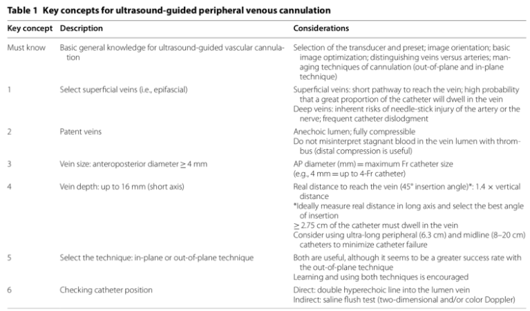
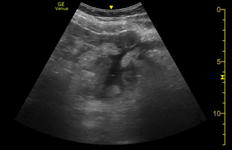
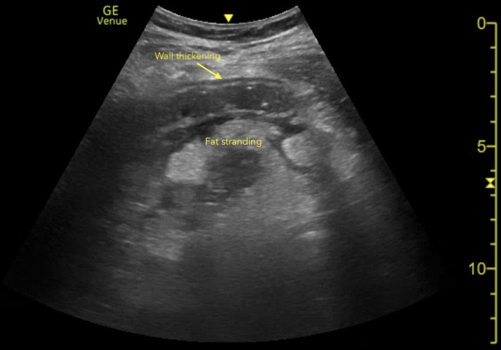
The first image is a transverse view of the uterus that shows free fluid in the retcouterine pouch (Pouch of Douglas). The second image is another transverse view of the uterus that also shows free fluid in the rectouterine pouch and then fans through to scan the uterus and adnexa. From what we see there is no gestational sac in the uterus and if you look closely there appears to be a heterogenous structure in the left adnexa. The final view is a FAST view in the right upper quadrant, looking at Morrison's Pouch. We see free fluid here as well. 

These findings - a positive pregnancy test, free fluid in the pelvis and no clear intra-uterine pregnancy indicates an ectopic pregnacny until proven otherwise. The next step should be a tranvaginal ultrasound and consultation with Gynecology. 

Conclusion and Learning Points:

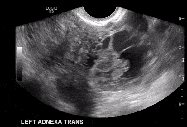
The transvaginal ultrasound revealed a left-sided ectopic pregnancy, as seen in the following picture. They identified a fetal pole and even a fetal heart rate in the ectopic pregnancy. The patient was taken to the operating room with Gynecology and had a salpingectomy without complications. She was discharged home three days later. 

Learning Points:

    • Any female of child-bearing age with abdominal pain should be considered for ectopic pregnancy
    • Ultrasound findings in ectopic pregnancy will not always show the ectopic itself, but rather findings suggestive of ectopic:
      • Intra-abdominal free fluid
      • No clear intra-uterine pregnancy (patients with ectopic will sometimes still have a "pseudo-gestational sac" that appears similar to a gestational sac, but there will be no yolk sac or fetal pole)
      • Heterogenous adnexal structure
    • You should not wait for B-HCG measurements to consider ectopic pregnancy, case reports have shown ectopic pregnancies with minimal HCG levels can still rupture (1)

References

1. Fu, Joyce, et al. Rupture of ectopic pregnancy with minimally detectable beta-human chorionic gonadotropin levels: a report of 2 cases. J Reprod Med. 2007 Jun;52(6):541-2.

This post was written by Charles Murchison MD and Anthony Medak MD, with further editing by Amir Aminlari MD.

Serratus Anterior Plane Block for Thoracic Wall Pain

Background


The serratus anterior plane block (SAPB) is a field block that provides analgesia from T2 to T9 dermatomes of the hemithorax by anesthetizing the lateral cutaneous branches of the intercostal nerves in that area. It was first described in a 2013 study demonstrating long-lasting thoracic-wall anesthesia with no significant adverse effects (1). It has since been utilized by anesthesia for prophylactic treatment of post-thoracotomy and post-mastectomy pain (2-4) and has demonstrated utility for treatment of rib fracture pain in the acute setting (5)

This case series describes its use in the Emergency Department (ED) for patients with thoracic wall pain from rib fractures, herpes zoster and chest tube placement. 

Serratus Anterior Plane Block in the Emergency Department: A Case Series.

Clinical Question

Is the SAPB feasible, safe and efficacious when used for a variety of thoracic wall pain syndromes in the ED?

Methods & Study Design

Case series in which six patients (age 60-94) underwent SAPB for treatment of thoracic pain.  Exclusion criteria were not specified; however, patients were included regardless of pre-procedural, multimodal analgesic use.  The authors recorded extent of injury and indication for SAPB. Outcomes of interest included efficacy of the nerve block and adverse events due to SABP during hospital stay.

SAPB was performed as follows: 25-30 mL of anesthetic (0.25% bupivacaine) was injected under ultrasound-guidance into the serratus anterior plane, either superficial (n=4) or deep (n=2) to the serratus anterior muscle. Sonographic landmarks for identification of the injection site included the lateral borders of the latissimus dorsi muscle and pectoralis muscle.

Image 1: Serratus anterior plane block sono-anatomy. Yellow line, target plane; purple-dotted line, needle; blue line, pleura. SCT, subcutaneous tissue; SAM, serratus anterior muscle; LDM, latissimus dorsi muscle; ICM, intercostal muscle (Lin et al 2020).

Results

SAPB was most commonly performed for pain related to anterior or posterior rib fractures (n=4), that was inadequately controlled with parenteral opioids.  SAPB provided complete or near-complete pain relief in these patients.  Additionally, SAPB resulted in significant pain relief for acute herpes zoster pain (n=1) and pre-procedural analgesia prior to tube thoracostomy (n=1).  Both superficial and deep injection locations resulted in effective analgesia.  No adverse events were noted.

Strength & Limitations

This is the first study to demonstrate efficacy of the SAPB for acute herpes zoster pain and procedural pain, and it adds to the growing body of literature supporting the use of SAPB for rib fracture pain. 

Limitations of this study include small sample size and lack of systematic data collection.  The authors note there is a possibility that physicians may not have documented all side effects.  Additionally, patients received non-standardized dosing of parenteral pain medication prior to SABP, therefore underdosing may have exaggerated the impact of the nerve block on pain relief.  Generalizability is limited as nerve blocks were performed by ultrasound fellowship-trained emergency physicians.

Authors Conclusion

“SAPB can be an effective analgesic modality for thoracic diseases and injuries including rib fractures, herpes zoster, and thoracostomy placement.”

Our Conclusion

SAPB was an effective adjunct to parenteral opioids in this case series.  Though limited, early data suggests that this is a safe and effective procedure.  Additional prospective studies are needed to compare SAPB to traditional techniques for the treatment of pain related to acute herpes zoster, thoracostomy, and rib fracture.

The Bottom Line 


Serratus anterior plane block, when performed by appropriately-trained physicians, is an effective and safe alternative to parenteral opioids and can provide significant, long-lasting analgesia for a variety of painful thoracic conditions. 

For more on the serratus anterior plane block see here:

Highland Ultrasound 


Authors

This post was written by Greta Davis, MS4 at UCSD School of Medicine, Charles Murchison, MD and Amir Aminlari MD. 

References

Lin J, Hoffman T, Badashova K, Motov S, Haines L. Serratus Anterior Plane Block in the Emergency Department: A Case Series. Clin Prac Cases Emerg Med. 2020 Feb;4(1):21-25.

1. Blanco R, Parras T, McDonnell JG, et al. Serratus plane block: a novel ultrasound-guided thoracic wall nerve block. Anaesthesia. 2013;68:1107-13.

2. Khalil AE, Abdallah NM, Bashandy GM, et al. Ultrasound-guided serratus anterior plane block versus thoracic epidural analgesia for thoracotomy pain. J Cardiothorac Vasc Anesth. 2017;31(1):152-8.

3. Rahimzadeh P, Imani F, Faiz SHR, et al. Impact of the ultrasound-guided serratus anterior plane block on post-mastectomy pain: a randomized clinical study. Turk J Anaesthesiol Reanim. 2018;46(5):388-92.

4. Madabushi R, Tewari S, Gautam SKS, et al. Serratus anterior plane block: a new analgesic technique for post-thoracotomy pain. Pain Physician. 2015;18(3):E421-4.

5. Durant E, Dixon B, Luftig J, et al. Ultrasound-guided serratus plane block for ED rib fracture pain control. Am J Emerg Med. 2017;35(1):197.e3-6.

Case 26: Genicular Nerve Block for Knee Pain – A Novel Technique

A 68 year old female with no significant past medical history presents to the Emergency Department (ED) with one day of right knee pain after falling off her bicycle onto her right side. She was immediately unable to bear weight on her right leg. 


Vitals: T 98.3, HR 73, RR 18, BP 114/70, SpO2 99%


Right leg exam: mild right knee effusion. No ligamentous laxity. Tenderness to palpation over lateral joint line > medial joint line. Tenderness to palpation over proximal anterior tibia. Knee extension limited due to pain. Neurovascularly intact with soft compartments.


Radiographic imaging demonstrated an isolated right tibial plateau fracture depression of the lateral plateau. The patient reports she is in severe pain but dislikes taking both over-the-counter and opioid pain medications. 

What nerves may be targeted to provide pain relief to her knee while maintaining motor function? What anatomic landmarks should be used on ultrasound to identify the branches of this nerve?

Answer and Learning Points

The genicular nerves derive from various major lower extremity nerve branches (femoral, obturator, sciatic, tibial) nerves and provide sensation to the knee capsule and joint. Cadaveric studies suggest that most genicular nerves are easily identifiable landmarks that may be used for therapeutic purposes. 5  Genicular nerve blocks (GNB) are traditionally used in this setting of chronic osteoarthritis knee pain via radiofrequency ablation or perioperative knee pain via ultrasound (1-4, 9).

The use of a GNBs in the ED is a novel technique to provide motor-sparing, pain relief for acute knee pain. This 68 year old patient with an isolated lateral tibial plateau fracture reported 4/10 pain over her proximal tibia at rest and 8/10 over her proximal tibia with movement. Written informed consent was obtained for GNBs of her right knee. Anatomic landmarks for the superior lateral (Image A,B) , superior medial (Image C,D), and inferior medial (Image D,E,F) genicular nerves were identified on ultrasound.

genicular nerve block

genicular nerve block

genicular nerve block

The ultrasound probe was placed in the sagittal orientation for each site. The superior lateral genicular nerve was located on ultrasound at the junction of the lateral femoral epicondyle and the epiphysis of the shaft of the femur, adjacent to the superior lateral genicular artery (Image A,B). The superior medial genicular nerve (SMGN) can be identified on ultrasound at the junction of the medial femoral epicondyle and the epiphysis of the shaft of the femur, adjacent to the superior medial genicular artery (Image C, D). The inferior medial genicular nerve (IMGN) can be identified on ultrasound at the junction of the medial tibial epicondyle and the epiphysis of the shaft of the tibia, adjacent to the inferior medial genicular artery (Image E, F, G) (6-8).

Under ultrasound guidance and using sterile technique, the skin was first anesthetized with 1% lidocaine after each site. A 21-gauge, 2 inch echogenic needle was inserted percutaneously and advanced under ultrasound guidance using an out-of-plate technique to inject 1.5 mL of 0.5% bupivacaine around the right superior lateral, superior medial, and inferior medial genicular nerves. 

Learning points

    • Genicular nerves derive from several lower extremity nerves and supply sensory innervation to the knee. 
    • The superior lateral, superior medial, and inferior medial genicular nerves are commonly targeted for pain relief with chronic knee osteoarthritis and postoperative pain.
    • The SLGN, SMGN, IMGN are easily located on ultrasound using anatomic landmarks (junction between epicondyles and epiphysis of the femur and tibia, adjacent to paired genicular arteries).
    • To obtain the images, you can use the linear probe in the sagittal location over lateral femoral epicondyle, medial femoral epicondyle, and medial tibial epicondyle.

References

1. Ahmed, Arif. “Ultrasound-guided radiofrequency ablation of genicular nerves of knee for relief of intractable pain from knee osteoarthritis: a case series.” British Journal of Pain, vol. 12, no. 3, 2017, pp. 145-154, https://www.ncbi.nlm.nih.gov/pmc/articles/PMC6058456/. Accessed 18 November 2020.

2. Caldwell, George L. “Reduced Opioid Use After Surgeon-Administered Genicular Nerve Block for Anterior Cruciate Ligament Reconstruction in Adults and Adolescents.” HSS Journal, vol. 15, no. 1, 2019, pp. 42-50, https://www.ncbi.nlm.nih.gov/pmc/articles/PMC6384209/. Accessed 18 November 2020.

3. Cankurtaran, Damla. “Comparing the effectiveness of ultrasound guided versus blind genicular nerve block on pain, muscle strength with isokinetic device, physical function and quality of life in chronic knee osteoarthritis: a prospective randomized controlled study.” Korean J Pain, vol. 33, no. 3, 2020, pp. 258 - 266, https://www.ncbi.nlm.nih.gov/pmc/articles/PMC7336352/. Accessed 18 November 2020.

4. Erdem, Yusuf. “The Efficacy of Ultrasound-Guided Pulsed Radiofrequency of Genicular Nerves in the Treatment of Chronic Knee Pain Due to Severe Degenerative Disease or Previous Total Knee Arthroplasty.” Med Sci Monit, vol. 25, 2019, pp. 1857 - 1863, https://www.ncbi.nlm.nih.gov/pmc/articles/PMC6423735/. Accessed 18 November 2020.

5. Fonkoué, Loïc. “Distribution of sensory nerves supplying the knee joint capsule and implications for genicular blockade and radiofrequency ablation: an anatomical study.” Surgical and Radiologic Anatomy, vol. 41, 2019, 1461–1471(2019), https://link.springer.com/article/10.1007/s00276-019-02291-y#citeas. Accessed 18 November 2020.

6. Güzelküçük, DemIr. “A different approach to the management of osteoarthritis in the knee: Ultrasound guided genicular nerve block.” Pain Medicine, vol. 18, no. 1, pp. 181 - 183, https://academic.oup.com/painmedicine/article/18/1/181/2924744. Accessed 18 November 2020.

7. Kim, Doo-Hwan. “Ultrasound-Guided Genicular Nerve Block for Knee Osteoarthritis: A Double-Blind, Randomized Controlled Trial of Local Anesthetic Alone or in Combination with Corticosteroid.” Pain Physician, vol. 21, 2018, pp. 41 - 51, https://www.ncbi.nlm.nih.gov/pmc/articles/PMC6058456/. Accessed 18 November 2020.

8. Protzman, Nicole. “Examining the feasibility of radiofrequency treatment for chronic knee pain after total knee arthroplasty.” PM&R, vol. 6, no. 4, 2014, pp. 373 - 376, https://pubmed.ncbi.nlm.nih.gov/24373908/. Accessed 18 November 2020.

9. Sahoo, Rajendra K. “Genicular nerve block for postoperative pain relief after total knee replacement.” Saudi J Anaesth, vol. 12, no. 2, 2020, pp. 235 - 237, https://www.ncbi.nlm.nih.gov/pmc/articles/PMC7164458/. Accessed 18 November 2020.

This post was written by Julia Sobel MD, with editing from Jessica Oswald MD, Charles Murchison MD and Amir Aminlari MD.

Erector Spinae Nerve Block for Abdominal Pain – A Case for Better Analgesia

Background


In the age of the opioid epidemic, there is a need for multi-modal pain control techniques, and nerve blocks will likely be an increasingly important piece of the puzzle, particularly in the Emergency Department. Ultrasound-guided nerve blocks for musculoskeletal complaints are now standard practice for many emergency physicians, so it is a reasonable next step to utilize this modality in lieu of opioids for abdominal complaints as well.

The erector spinae plane (ESP) block has been shown to improve pain control for rib fractures in the emergency department, and a more inferior approach has demonstrated success in the perioperative period for abdominal surgeries (1,2). However, this type of block has not been studied for the management of abdominal pain in the emergency department.

This study examined the efficacy of the ESP block on patients with acute appendicitis to see if it could reduce opioid use.


A Novel Technique to Reduce Reliance on Opioids for Analgesia from Acute Appendicitis: The Ultrasound-guided Erector Spinae Plane Block

Clinical Question

Can an inferiorly located, ultrasound-guided erector spinae plane block successfully manage the pain of acute appendicitis in the emergency department setting?

Methods & Study Design

Case report in which ultrasound guidance was used to perform an ESP block at the L1 level.

The patient was a 24-year-old male with uncomplicated appendicitis as diagnosed on CT. The location for needle insertion was identified by palpating the L1 spinous process and placing a linear probe 3 cm lateral, at the transverse process. A Touhy needle was advanced under ultrasound guidance to the level of the transverse process, and saline hydrodissection was used to confirm the needle tip in the fascial plane. Then, 20mL of 1% lidocaine was injected.

Results

Initially, the patient was reporting 7/10 pain following analgesia with 0.5 mg IV hydromorphone, 30 mg IV ketorolac, and 1 g IV acetaminophen. Thirty minutes following placement of the erector spinae plane block, the patient reported 0/10 pain without palpation, and 3/10 pain with deep palpation. Testing to cold revealed loss of sensation between the T10-L2 dermatomes. The patient did not require additional analgesia during the rest of his 5.5-hour emergency department stay. 

Strength & Limitations

While this is a case study of only one patient, it provides explicit guidance on performing an erector spinae plane block in the emergency department setting. However, the primary method of assessing pain control is inherently subjective and may be limited by the patient’s perception of the efficacy of the block. Further, the patient had already received analgesia, which may have impacted the efficacy of the block over the course of the ED stay. 

Authors Conclusion

A single injection, ultrasound-guided erector spinae plane block can provide complete analgesia for appendicitis.

Our Conclusion

Performing an erector spinae plane block at the L1 level was an effective adjunct to opioid analgesia in this case. Because of the relative safety of this block and efficacy in this case, it warrants further investigation as to the ideal level for pain control, particularly in a larger sample of patients. 

The Bottom Line 


When performed in a more inferior position, an ultrasound-guided erector spinae plane block may be an effective form of analgesia for appendicitis in the emergency department setting. 

For excellent guides on how to perform erector spinae blocks see here:

EMRA

Highland Ultrasound 

Authors

This post was written by Kaley Waring, MS4 at UCSD School of Medicine, Charles Murchison MD and Amir Aminlari MD.

References

Mantuani et al. A Novel Technique to Reduce Reliance on Opioids for Analgesia from Acute Appendicitis: The Ultrasound-guided Erector Spinae Plane Block. Clin Pract Cases Emerg Med. 2019 Aug; 3(3): 248–251.

1. Luftig J, Mantuani D, Herring AA, et al. Successful emergency pain control for posterior rib fractures with ultrasound-guided erector spinae plane block. Am J Emerg Med. 2018;36(8):1391–6

2. Chin KJ, Adhikary S, Sarwani N, et al. The analgesic efficacy of pre-operative bilateral erector spinae plane (ESP) blocks in patients having ventral hernia repair. Anaesthesia. 2017;72(4):452–60

Case 25: Aortic Dissection

A 44 year old male with a history of heroin abuse presents to the emergency department with altered mental status.  Per EMS, the patient was found on the street with decreased level of consciousness and poor respiratory effort.  EMS was concerned about opioid overdose, and he was treated with 4mg Narcan, with improvement in his mental status.  

Upon arrival to the ED, he was noted to be agitated and tachypneic with RR in the 40’s.  

Vitals: BP 90/65 mmHg, HR 110, O2 100% on RA, glucose 158.

He is alert and oriented to person, month, and place, but appears agitated and confused.  He denies any complaints other than shortness of breath, and states he felt fine before using heroin.  He denies any past medical history.

Exam notable for tachycardia, diffuse rhonchi throughout all lung fields, 2+ nonpitting lower extremity edema.  He is neurologically intact with 2+ pulses throughout.

A bedside echocardiogram was performed, what do you see?  What are your next steps?

PS long
dilated aortic root
aortic dissection suprasternal ultrasound
abdominal aortic dissection ultrasound
dissection to iliacs ultrasound

Answer and Learning Points

Answer:

The first two images show a parasternal long-axis view, with a dilated aortic root measuring approximately 4.2cm.  Also notice the pericardial effusion with a homogenous layer that appears fixed to the pericardium.  At the time it was unclear whether this represented a clot within the pericardial sac, or was superficial to it.

Given the dilated aortic root, a suprasternal short-axis view was obtained to assess the proximal aorta, with the short axis view seen on the third image.  A flap was visualized in the aortic lumen, significant for an ascending aortic dissection.  The dissection was then visualized in the abdominal aorta, extending distal to the common iliac arteries, seen in the last images.

The patient was placed on esmolol drip and later required vasopressor support.  CT angiography was obtained, confirming a Type A aortic dissection.  He was transferred to a nearby hospital for emergent repair of his ascending and descending aortic dissection.  

Per the operative report, the patient had developed a significant pericardial effusion by the time he reached the OR, and the visualized homogenous layer above most likely represented a blood clot within the pericardium.

Learning Points:

    • While CTA remains the gold standard for diagnosis of aortic dissection, POCUS remains a great tool for diagnosing both ascending and descending aortic dissection, particularly in the unstable patient.
    • In addition to directly visualizing the dissection flap, TTE can also be used to identify patients with high risk features, such as those with cardiac tamponade, severe aortic dilatation, severe aortic regurgitation, regional wall motion abnormalities, and decreased ejection fraction (1).
    • TTE has been shown to have a sensitivity of 78-90% and specificity 87-96% for type A dissection in older studies (2,3), and in more recent studies showing improved sensitivity up to 97-99% (1,4,5) and specificity 100% (4) with improved image quality.
    • The suprasternal notch views are particularly useful in evaluating the proximal ascending aorta, and allow the operator to assess for aortic dissection, coarctation, dilatation of the aortic arch, and retrograde flow from the descending aorta.

References

1. Sobczyk D, Nycz K. Feasibility and accuracy of bedside transthoracic echocardiography in diagnosis of acute proximal aortic dissection. Cardiovasc Ultrasound. 2015;13:15.

2. Evangelista A, Flachskamp FA, Erbel R, Antonini-Canterin F, Vlachopoulos C, Rocchi G, et al. Echocardiography in aortic diseases: EAE recommendations for clinical practice. Eur J Echocardiogr. 2010;11:645–58. doi: 10.1093/ejechocard/jeq056.

3. Nienaber CA, von Kodolitsch Y, Nicolas V, Siglow V, Piepho A, Jaup T, et al. The diagnosis of thoracic aortic dissection by noninvasive imaging procedures. N Engl J Med. 1993;328:1–9. doi: 10.1056/NEJM199301073280101.

4. Cecconi M, Chirillo F, Constantini C, Iacobone G, Lopez E, Zanoli R, et al. The role of transthoracic echocardiography in the diagnosis and management of acute type A aortic syndrome. Am Heart J. 2012;163(1):112–8. doi: 10.1016/j.ahj.2011.09.022.

5. Nazerian, P., Vanni, S., Castelli, M. et al.Diagnostic performance of emergency transthoracic focus cardiac ultrasound in suspected acute type A aortic dissection. Intern Emerg Med9, 665–670 (2014). https://doi.org/10.1007/s11739-014-1080-9

This post was written by Rachna Subramony MD, Alex Anshus MD, with editing from Sukhdeep Singh MD, Charles Murchison MD and Amir Aminlari MD.

A Practical Guide to Placing Ultrasound-Guided Peripheral IVs

ultrasound iv

Background


Peripheral IV placement is one of the most common procedures performed in hospitals, with hundreds of millions performed each year. For the most part, IVs are successfully placed in the traditional way - using landmarks and visualization/palpation of the vessels. But as we all know, there are several patient factors that can make peripheral IV placement more difficult, including obesity, edema, a history of IV drug use, sickle cell disease, lupus, diabetes, etc.

Ultrasound-guided IV placement is increasingly used in the emergency room, medicine floors and ICUs in patients with difficult IV access, but  the research is clear that there is a higher premature failure rate with ultrasound-guided IVs (1). 

This practical guideline details several considerations that may help reduce the premature failure rate of ultrasound-guided IV catheter placement.  


Ultrasound-Guided Peripheral Venous Cannulation in Critically Ill Patients: a Practical Guideline

Clinical Question

What are key concepts to help reduce ultrasound guided peripheral IV catheter complications, prolong life of catheters, and increase rate of successful placement?

Methods & Study Design

• Design 

Review paper

Results

There are six key concepts to help minimize complications and increase duration of ultrasound-guided peripheral IV catheter placement.

Strength & Limitations

Potential limits to these guidelines include ultrasound experience level of a person placing IV catheters, whether the necessary equipment is routinely available at lower resource centers, and the setting in which IV cannulation takes place (e.g. trauma or non-trauma). The access and cost of ultra-long peripheral and midline catheters may limit use given potential for high utilization. 

Authors Conclusion

Practitioners should consider several issues when inserting intravenous peripheral catheters under ultrasound guidance, aiming to improve success rate, avoid complications and lengthen the survival of the catheter. Based on available data and everyday practice, all indicate that catheters longer than standard size are needed for US-guided peripheral venous cannulation, with the purpose of minimizing premature catheter failure. This is a call for attention to catheter manufacturers, since a more affordable solution at hand is expected from them shortly.”

Our Conclusion

For placement of ultrasound-guided peripheral IVs consider these rules:

    • Always use a long IV catheter, preferably 6 cm or longer
    • Choose veins that are:
      • At least 4 mm in diameter
      • At most 1.5 cm deep
      • As distal as possible, preferably distal to the antecubital fossa
    • At last 2.75 cm of the catheter should be in the vein
    • Check IV placement by flushing saline and use the ultrasound to watch a proximal vessel for turbulent flow

The Bottom Line 

When identifying an appropriate vein for ultrasound guided IV access, choose a superficial, patent, large, distal vein to minimize distance needed for the catheter to travel. Ensure adequate catheter length and confirm catheter position after placement to decrease failure rate.

Authors

This post was written by Tori Speck, MS4 at UCSD School of Medicine, Charles Murchison, MD and Amir Aminlari, MD. 

References

Blanco, Pablo. “Ultrasound-Guided Peripheral Venous Cannulation in Critically Ill Patients: a Practical Guideline.” The Ultrasound Journal, Springer Milan, 17 Oct. 2019, www.ncbi.nlm.nih.gov/pubmed/31624927. 

1. Bahl, Amit, et al. “Ultralong Versus Standard Long Peripheral Intravenous Catheters: A Randomized Controlled Trial of Ultrasonographically Guided Catheter Survival.” Annals of Emergency Medicine, Mosby, 16 Jan. 2020, www.sciencedirect.com/science/article/pii/S0196064419313836. 

2. Gottlieb, Michael et al. “Comparison of Short- vs Long-axis Technique for Ultrasound-guided Peripheral Line Placement: A Systematic Review and Meta-analysis.” Cureus vol. 10,5 e2718. 31 May. 2018, doi:10.7759/cureus.2718

3. Presley, Brad. “Ultrasound Guided Intravenous Access.” StatPearls [Internet]., U.S. National Library of Medicine, 31 July 2020, www.ncbi.nlm.nih.gov/books/NBK525988/.

Will Ultrasound Help Your Success with Arterial Lines?

radial a line

Background


Traditionally, we are taught to place radial artery catheters - A lines - using anatomy and pulse palpation. First-time success rate varies in the literature from as low as 15% to about 70%, with complications including hemorrhage or hematoma. Patients with particularly challenging insertions include small children, as well as adults with hypotension, obesity, or peripheral edema.

Improved procedural success rates, safety and cost effectiveness using ultrasound guidance has been demonstrated extensively in central venous catheterization, however, this has not yet been established for arterial catheterization, as literature for US guided radial artery catheterization has been both limited and presents conflicting results.

Efficacy of ultrasound-guided radial artery catheterization: a systematic review and meta-analysis of randomized controlled trials. 

Clinical Question

Is US guidance of radial artery catheterizations effective compared to standard methods of palpation/Doppler in either adult or pediatric patients?

Methods & Study Design

• Design 

Systematic review and meta-analysis 

• Population 

Adult or pediatric patients requiring radial artery catheterization, inclusion criteria varied by study. 

• Intervention 

Ultrasound-guided radial artery catheterization compared to doppler-assisted or landmark technique.

• Outcomes  

First attempt success rate and complications from attempts at radial artery catheterization.

Results

7 RCTs were used to calculate a pooled estimate of first-attempt success

    • Rate of first-attempt success in US group: 48.5%
    • Rate of first-attempt success in control group: 30.7%
    • US-guided radial arterial catheterization was associated with increased first-attempt success (RR 1.55, 95% CI, 1.02 to 2.35, P = 0.04)
    • US-guided radial arterial catheterization significantly reduced mean attempts to success (WMD −1.13, 95% CI −1.58 to −0.69, P <0.001), mean time to success (WMD −72.97 seconds, 95% CI −134.41 to −11.52, P = 0.02), and incidence of hematoma (RR 0.17, 95% CI 0.07 to 0.41, P <0.001)

Subgroup Analyses

    • No difference in primary outcome between elective insertion (five trials, RR 1.91, 95% CI, 1.45 to 2.53) and emergency insertion (two trials, RR 1.05, 95% CI, 0.38 to 2.83) 
    • US-guided radial arterial catheterization was associated with significantly increased first-attempt success in small children/infants (RR 1.94, 95% CI, 1.31 to 2.88, P = 0.001)

Strength & Limitations

Strengths:

Well-performed systematic review and meta-analysis with a clear primary outcome and relevant secondary outcomes.

Limitations:

    • There were major differences in ultrasound experience of operators, ranging from those without any experience and only an observational training period to expert operators.
    • Overall, the studies had small sample sizes and only a small number of studies met inclusion criteria for the review.
    • Not enough samples to conduct additional subgroup analyses of patients who might be characterized as difficult-to-insert, including hypotension, obesity, edematous, and pulseless.
    • Lack of inclusion of other potential factors including patient pain or patient/operator satisfaction.
    • Lack of comment and description on specific US-guidance techniques


Authors Conclusion

"US guidance is an effective and safe technique for radial artery catheterization, even in small children and infants. However, results should be interpreted cautiously due to the heterogeneity among studies."

Our Conclusion

Though data from RCTs is limited, with proper operator training US-guidance can be an effective method of improving radial artery catheterization accuracy, especially in small children/infants with smaller and more difficult-to-palpate anatomy.

Conflicting results in previously conducted RCTs may be attributed to differences in operator training or lack of a proper observational training period and thus careful consideration of the operator should be conducted in future RCTs. A physician who has performed dozens of A-lines using the palpation technique, who is unfamiliar with bedside ultrasonography, is unlikely to benefit significantly from adding this modality to their procedure, while residents who are trained with ultrasounds in their hands will likely benefit more. 

Future RCTs should focus on patient populations that have been characterized as difficult-to-insert including hypotension which is in particular significant to emergency medicine, as well as edematous or obese patients.

The Bottom Line 

Ultrasound-guided placement of radial artery catheters is effective compared to standard palpation techniques, and should be taught to current Emergency Medicine residents. Further studies are needed to elucidate the effect of US on difficult-to-cath patients, as the effect is hypothesized to be magnified in patients who are hypotensive or edematous.

Authors

This post was written by Jessica Wen, MS4 at UCSD School of Medicine, Charles Murchison, MD and Amir Aminlari, MD. 

References

Gu W, Tie H, Zeng X. Efficacy of ultrasound-guided radial artery catheterization: a systematic review and meta-analysis of randomized controlled trials. Critical Care. 2014; 18(3): R93 doi:10.1186/cc13862

Case 24: Diverticulitis


A 56 year old male with a history of uncomplicated diverticulitis presented to the emergency room with left lower quadrant pain and loose stools for the last six days. He denies fever, vomiting or blood in hist stool 

Vitals: T 97.3   BP 152/81   HR 91       RR 18      SPO2 97% on RA


You physical exam shows tenderness to palpation in the left lower quadrant with no peritoneal signs. You are on the fence about getting a CT abdomen and pelvis with contrast to look for an abscess versus treating this as uncomplicated diverticulitis. You decide to throw the ultrasound probe on the area of his pain. What do we see in these images? How would this change management?


Answer and Learning Points

Answer:

The three videos and two images show diverticulitis with an abscess or phlegmon beneath the bowel loops. Though CT is the gold standard for diagnosing diverticulitis, ultrasound is relatively sensitive in the diagnosis and has the advantage of being cheap, fast and radiation-free (1). 

When looking for diverticulitis on ultrasound physicians will typically use a "lawn mower" approach to the left abdomen to search for areas of affected bowel. One way to get to the area of interest more quickly is simply ask the patient to point to the area of maximal tenderness and start there, similar to appendicitis or small bowel obstruction. There are a few findings on ultrasound that indicate diverticulitis (2,3):

  1. Thickening of bowel wall, typically at least 4-5mm
  2. Echogenic fat surrounding the bowel, which is representative of fat stranding seen on CT
  3. Diverticulum
diverticulitis ultrasound
Wall thickening and fat stranding


Ultrasound is also helpful in looking for abscess, such as in our case. We see there is an area of hypoechogenicity with no color flow, representing likely abscess adjacent to the bowel.  


Our patient ultimately got a CT scan that confirmed he had diverticulitis with abscess. He was admitted to medicine with GI and surgery consults following.

References

(1) Lameris, W et al. Graded compression ultrasonography and computed tomography in acute colonic diverticulitis: meta-analysis of test accuracy. Eur Radiol. 2008 Nov;18(11):2498-511.

(2) Schwerk, WB et al. Sonography in acute colonic diverticulitis. A prospective stud. Dis Colon Rectum. 1992 Nov;35(11):1077-8

(3) Mazzei M et al. Sigmoid diverticulitis: US findings. Crit Ultrasound J. 2013 Jul 15;5 Suppl 1(Suppl 1):S5.

This post was written by Charles Murchison MD, with editing from Colleen Campbell MD and Amir Aminlari MD.

Translate »