

Background
Necrotizing fasciitis (NF) is rapidly progressing, severe soft tissue infection with a mortality rate of 19.3% with treatment and significantly higher without treatment (1). Early diagnosis is essential to prompt surgical intervention and reduce morbidity and mortality. However, treatment can often be delayed because no laboratory or imaging test can definitively diagnose NF. Contrast-enhanced CT shows the best accuracy, but again is not perfect and can be difficult to obtain in unstable patients. MRI is similarly accurate, but even less feasible in the Emergency Department. Ultimately, it remains a surgical diagnosis.
Ultrasonography is a rapid, bedside, and non-invasive tool that has potential to accelerate assessment of patient with clinical suspicion for NF. There are ultrasonographic findings associated with NF diagnosis, including irregularity or thickening of deep fascia, subcutaneous emphysema, and fluid accumulation along the deep fascial plane (2-13). Considering this condition’s rapid progression, ultrasonography may enable physicians to quickly gauge disease severity and triage accordingly, prompting earlier surgery and bettering patient outcomes.
Clinical Question
What is the relationship between ultrasonographic finding of fluid accumulation along the deep fascia and diagnosis and prognosis of necrotizing fasciitis?
What ultrasonographic findings are significantly different between NF patients and non-NF patients?
What is the ultrasonographic-detected depth of fluid accumulation along the deep fascia that offers the greatest accuracy to diagnosis of NF?
Is there a difference in the prognosis between NF patients with fluid accumulation compared to NF patients without fluid accumulation?
Methods & Study Design
• Design
Retrospective study with prospective enrollment
• Population
This study was conducted at Chang Gung Memorial Hospital, a suburban academic tertiary care hospital.
Inclusion criteria: patients who visited the ED from February 2015 – November 2016 with clinical suspicion of NF of limbs based on symptoms and clinical signs (severe pain out of proportion, skin findings, rapid progression, crepitus, skin bullae, necrosis, or ecchymosis).
NF group: discharge diagnosis of NF, confirmed by pathology report showing necrosis after surgical intervention
Non-NF group: did not have surgical intervention or whose pathology report did not support NF diagnosis
Exclusion criteria: patients with ED visits between 24:00 – 7:00, non-lesion side also has fluid accumulation, age <18yo, prior antibiotics or debridement, lesions involving trunk area
• Intervention
Ultrasonographic exam within 1 hour after ED arrival completed by one of three experienced emergency physicians who received an 8-hour basic and soft-tissue ultrasonographic training before the study
Orthopedic consult for surgical opinion
• Outcomes
- Diagnostic markers: irregularity or thickening of deep fascia, fluid accumulation, subcutaneous emphysema, subcutaneous cobblestone
- Reasonable cutoff value of fluid accumulation along deep fascial plane for diagnosing NF according to receiving operating characteristic (ROC) curve
- Prognostic markers: length of stay (LOS) in hospital, mortality, amputations, number of operations
Results
Ultrasound finding of fluid accumulation and irregular or thickened fascial layer were significantly different between NF and non-NF groups. All patients who had subcutaneous emphysema were in the NF group.
The best cutoff point of fluid accumulation to diagnose NF was 2mm, which had the best accuracy (72.7%), with sensitivity of 75%, a specificity of 70.2%, a positive predictive value of 71.7% and a negative predictive value of 72.7%.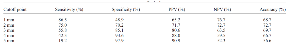
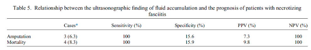
NF patients with fluid accumulation had longer length of stay than NF patients without fluid accumulation (average: 39 days vs. 23 days). Number of operations were not significantly different between NF patients with and without fluid accumulation. All NF patients who had an amputation or died had fluid accumulation.
Overall mortality between NF and non-NF groups showed no significant difference.
Strength & Limitations
Strengths
- Sample size was larger than other studies investigating ultrasonographic findings for NF diagnosis.
- Study had a comparator groups with clear definitions (NF vs. non-NF).
- Ultrasound training was standardized and assessed with inter-rater reliability between three emergency physicians as 100%.
Limitations
- Small, imbalanced sample of NF patients for sensitivity and specificity analysis of fluid accumulation for amputation and mortality.
- Study excluded patients with truncal soft tissue infections.
- Study excluded patients with prior antibiotics or debridement, which may have been NF patients with higher severity and worse prognosis.
- Patient population were from south Taiwan exclusively.
- NF patients had higher prevalence of specific co-morbidities (diabetes mellitus, liver cirrhosis, and alcohol use disorder), which could be confounding.
Authors Conclusion
“The ultrasonographic finding of fluid accumulation along the deep fascia with a cutoff point of more than 2 mm of depth may aid in diagnosing NF. For the prognosis of NF, when fluid accumulation was present along deep fascia on ultrasound, patients with NF had longer lengths of hospital stays and were at risk of amputation or mortality. Ultrasonography is a point-of-care imaging tool that facilitates the diagnosis and prognosis of NF.” (14)
Our Conclusion
Consistent with prior studies and case reports (2-13), this study supports the role of ultrasound in the diagnosis of NF. Trained emergency physicians were able to successfully use ultrasound to detect significant imaging differences in NF patients, including fascial irregularity and deep fascial fluid accumulation. In comparison to Yen et al., this study suggests an even lower cutoff point of fluid accumulation along the deep fascia (2mm vs 4mm) for the highest diagnostic accuracy. We would caution that the finding of "fluid accumulation" was somewhat difficult to interpret in their study.
Further studies with larger sample sizes need to be completed. However, with the diagnostic and prognostic trends seen in this study, ultrasound should be considered as a timely, efficient imaging modality that can help identify patients with clinical suspicion of NF and accelerate OR intervention.
The Bottom Line
Ultrasound is a viable imaging modality for patients with clinical suspicion of NF that could potentially expedite surgical intervention, though imaging findings may not be as easy to interpret as the authors lay out.
Authors
This post was written by Caresse Vuong, Charles Murchison MD and Amir Aminlari MD.
References
- Khamnuan P, Chongruksut W, Jearwattanakanok K, Patumanond J, Yodluangfun S, Tantraworasin A. Necrotizing fasciitis: Risk factors of mortality. Risk Manag Healthc Policy 2015;8:1–7.
- Castleberg E, Jenson N, Am Dinh V. Diagnosis of necrotizing fasciitis with bedside ultrasound: The STAFF exam. West J Emerg Med 2014;15:111–113.
- Tsai CC, Lai CS, Yu ML, Chou CK, Lin SD. Early diagnosis of necrotizing fasciitis by utilization of ultrasonography. Kaohsiung J Med Sci 1996;12:235–240.
- Wronski M, Slodkowski M, Cebulski W, Karkocha D, Krasnodebski IW. Necrotizing fasciitis: Early sonographic diagnosis. J Clin Ultrasound 2011;39:236–239.
- Yen ZS, Wang HP, Ma HM, Chen SC, Chen WJ. Ultrasonographic screening of clinically-suspected necrotizing fasciitis. Acad Emerg Med 2002;9:1448–1451.
- Bernardi, Emanuele, Antonello Iacobucci, Letizia Barutta, Elisa Pizzolato, Virna Olocco, and Bruno Tartaglino. “A-Lines in Necrotizing Fasciitis of the Lower Limb.” Journal of Ultrasound in Medicine 33, no. 11 (2014): 2044–46.
- Chao, H. C., M. S. Kong, and T. Y. Lin. “Diagnosis of Necrotizing Fasciitis in Children.” Journal of Ultrasound in Medicine: Official Journal of the American Institute of Ultrasound in Medicine 18, no. 4 (April 1999): 277–81.
- Hosek, William T., and Timothy C. Laeger. “Early Diagnosis of Necrotizing Fasciitis with Soft Tissue Ultrasound.” Academic Emergency Medicine 16, no. 10 (2009): 1033–1033.
- Oelze, Lindsay, Stanley Wu, and Jennifer Carnell. “Emergency Ultrasonography for the Early Diagnosis of Necrotizing Fasciitis: A Case Series from the ED.” The American Journal of Emergency Medicine 31, no. 3 (March 1, 2013): 632.e5-632.e7.
- Kehrl, Thompson. “Point-of-Care Ultrasound Diagnosis of Necrotizing Fasciitis Missed by Computed Tomography and Magnetic Resonance Imaging.” The Journal of Emergency Medicine 47, no. 2 (August 2014): 172–75.
- Shyy, William, Roneesha S. Knight, Ruth Goldstein, Eric D. Isaacs, and Nathan A. Teismann. “Sonographic Findings in Necrotizing Fasciitis.” Journal of Ultrasound in Medicine 35, no. 10 (2016): 2273–77.
- Hanif, Muhammad A., and Michael J. Bradley. “Sonographic Findings of Necrotizing Fasciitis in the Breast.” Journal of Clinical Ultrasound: JCU 36, no. 8 (October 2008): 517–19.
- Valle Alonso, Joaquín, Ganapathiram Lakshmanan, and Yasser Saleem. “Use of POCUS Ultrasound in Sepsis, Bedside Diagnosis of Necrotizing Fasciitis.” QJM: An International Journal of Medicine 110, no. 10 (October 1, 2017): 687–88.
- Lin, Chun-Nan, Cheng-Ting Hsiao, Chia-Peng Chang, Tsung-Yu Huang, Kuang-Yu Hsiao, Yi-Chuan Chen, and Wen-Chih Fann. “The Relationship Between Fluid Accumulation in Ultrasonography and the Diagnosis and Prognosis of Patients with Necrotizing Fasciitis.” Ultrasound in Medicine & Biology 45, no. 7 (2019): 1545–50.